Practical work on microbiology design. Workshop on veterinary microbiology
INSTITUTIONS OF HIGHER EDUCATION
V. N. Kislenko
Workshop
By veterinary
Microbiology
And immunology
Committed by the Ministry of Agriculture of the Russian Federation in
quality tutorial for higher students
educational institutions students in the specialty
"Veterinary"
Moscow "Koloss" 2005
UDC 619: 579 (075.8) BBK 48Я73 K44
Editor TS MYOCHEVA
Reviewers: Doctor of Veterinary Sciences, Professor V. I. Pleshakov(Institute Vetee
rinar medicine Omsk GAU); Doctor of Veterinary Sciences, Professor IT.. I. Ba.
ruvennikov(Institute of Veterinary Medicine Altai GAU)
Kislenko V.N.
K44 Workshop on veterinary microbiology and immunology. - M.: Colossus, 2005.- 232 p. L.: Il. - (textbooks and studies. Manuals for students Higher. Studies. Institutions). ISBN 5-9532-0332-2
The workshop consists of two sections. The "General Microbiology" section contains information on the rules of operation in the bacteriological laboratory, a description of the main microbiological, genetic and immunological methods for the study of microorganisms. In the "Infectious Diseases" section Methods listed laboratory diagnostics, Differentiation of pathogens and a list of used biological products.
Methodical guidelines for practical training for teachers are given.
Kits of tests on electronic media (CD-disk) are attached in general and private microbiology, immunology, as well as by theoritical course.
For students of higher educational institutions in the specialty "Veterinary".
Introduction
Veterinary laboratories are institutions of the state veterinary service, the activities of which are aimed at ensuring well-being in animal husbandry, the prevention and liquidation of diseases and death of animals, as well as to protect the population from diseases common to animals and human. By appointment, veterinary laboratories are district, interdistrict (zonal), regional (boundary) and republican.The main task of veterinary laboratories is to establish an accurate diagnosis of diseases of farm animals, including birds, fur animals, fish and bees, as well as conducting examination of meat, milk and other food products of animal and vegetable origin. In laboratories also perform scientific work, produce the production of some biostimulants, antibiotics, etc.
The material for laboratory research is blood, urine, sputum, milk, feces, the contents of the abscesses (PM) obtained during the life of the animal; Piens of parenchymal organs or other fabrics after their death, samples of environmental objects (water, air, soil, feed, plants, washes with care objects). The material in the laboratory is investigated by bacteriological, serological, histological, biochemical, micrological, and toxicological methods, for which the necessary conditions must be created (specially designated premises, equipment, microclimate, etc.).
The laboratory occupies a separate building, the location away from the passage roads. It should contain a reception office, Pato-loganoic, bacteriological, serological, biochemical and virological departments, as well as special containers for thermostats, washing dishes, autoclave. In the dishwashing room there are tables, sinks, hot and cold water supply, gas or electric stove, shelving for washed dishes, exhaust cabinet, enameled bathtubs, basins and other tanks, a solution of acid in glass vessels for disinfection of pipettes, slim glasses and other dishes . A separate room is discharged to bacteriological cuisine (medium-rich), where the nutrient media is prepared for the cultivation of microorganisms, prepare dishes for sterilization. Here in the cabinets store sterile dishes and well-packed chemicals, nutrient components.
To perform work in aseptic conditions, equip special insulated rooms - boxes(English Box - box), consisting of boxing itself and pre-kiss. Desktop boxes, items and air are used in which they are disinfected with WFA lamps, and laminar cabinets, where the active removal of air is used.
Laboratory animals (white mice, guinea pigs, white rats, rabbits, etc.) are placed in vivaria.As a rule, in Vivaria also contain healthy donor rashes, the blood is used to react complement (RSK) and the preparation of nutrient media. Infected laboratory animals contain in an isolated room.
In addition, there are rooms for specialists serving staff, head office, library, weight, changing room, warehouse, etc.
To maintain proper cleanliness, the floor in the rooms are covered with linoleum or tiled tiles. Walls and ceilings, as a rule, smooth (without eaves and stucco decorations), with rounded corners, painted in bright tones of oil paint. Ceilings can be shaving lime. It is desirable to bind the walls with plastic or tiled from the floor to the ceiling.
In the laboratory there must be hot and cold water, sewage, pedal buckets for garbage, which are released daily, wash and disinfect, towels, soap and disinfecting solution. In the rooms there is only the most necessary equipment: tables, cabinet for storing small equipment, paints, reagents, dishes, tools, etc. Tables are usually installed in front of the windows. They must be stable, comfortable, 80 cm high, with a tip. The surface of the tables is covered with plastic or linoleum, glass or white special paint. The table places a microscope, as well as the necessary items for bacteriological work.
The bacteriological research method, as a rule, includes microscopy, isolating and studying the properties of a clean culture of the causative agent of the disease and infection of laboratory animals (biological sample). The results of bacteriological analysis by signature of the head of the department or director of the laboratory report only to officials: a veterinary doctor, zoo-engineer, to the head of the enterprise.
Microbiological laboratory equipment.
The following devices and devices are needed to work in the laboratory: biological immersion microscopes with additional devices (illuminator, phase-contrasting device, nonopoly condenser, etc.), fluorescent microscopes, thermostats, equipment for sterilization, pH meters, devices forreceipt of distilled water (distiller), centrifuge, technical and analytical scales, filtering equipment (Zathetz et al.), Water baths, refrigerators, machine for making cotton-gauze plugs, a set of tools (bacteriological loops, spatulas, needles, tweezers and Dr.), Laboratory dishes (test tubes, flasks, Petri dishes, mattresses, bottles, ampoules, parser and graduated pipettes), etc.
The laboratory highlighted a special place for coloring microscopic drugs, where there are solutions of special dyes, alcohol, acid, filtering paper, etc. each workplace Equipped with a gas torch or alcohol, a jar with a disinfectant solution. For daily work The laboratory should have the necessary nutrient media, chemical reagents, diagnostic preparations and other laboratory materials. In large laboratories there are thermostat rooms for mass cultivation of microorganisms, producing serological reactions.
For growing, storage of crops, sterilization of laboratory dishes and other purposes, the following equipment is intended:
Thermostat. The device in which the constant temperature is supported. The optimal temperature for the reproduction of many microorganisms is 37 "C. Thermostats are air and water.
Microanostat. Apparatus for growing microorganisms in anaerobic conditions.
Refrigerators. Used in microbiological laboratories for storing crops of microorganisms, nutrient media, blood, sera, vaccines and other biological preparations at a temperature of about 4 ° C. For storage of preparations below 0 ° C, low-temperature refrigerators are designed, in which the temperature -20 "C and below is supported.
Centrifuges. Used to precipitate microorganisms, erythrocytes and other cells, separation of inhomogeneous liquids (emulsions, suspension). In laboratories, centrifuges with different modes of operation are used.
Drying-sterilization cabinet (pasteur furnace). Designed for air sterilization of laboratory dishes and other materials.
Steam sterilizer (autoclave). Designed for sterilization by ferry under pressure. In microbiological laboratories, autoclaves of different models are used (vertical, horizontal, stationary, portable).
Rules of work in the microbiological laboratory.
The microbiologist deals mainly with pure cultures of microorganisms, which are the offspring of one cell. Since in the air and on the surface of items in the laboratory (on the tables, devices, instruments, as well as on clothing, etc.) there are always many diverse microorganisms, one should constantly take care of the preservation of the purity of the cultures being studied. Therefore, when working in microbiological laboratory It is necessary to strictly follow certain rules, one of which is to maintain purity, including daily hygienic cleaning of all rooms.To destroy microorganisms in the air and on surfaces there are various disinfection methods.
Air in the laboratory is partially purified by ventilating. Ventilation sharply reduces the number of microorganisms in the air, especially with a significant temperature difference outside and indoors. The duration of the ventilation is at least 30 ... 60 min.
The more efficient and most commonly used method of disinfection of air is the effect of ultraviolet radiation (WFIC), which has a high antimicrobial effect and causing the death of not only vegetative cells, but also a dispute of microorganisms. Due to the weak penetrating ability, ultraviolet radiation does not pass through ordinary glass and is easily absorbed by dust particles. Therefore, for sterilization, irradiation time is from 30 minutes to several hours, depending on the degree of air pollution.
As a source of UFI, bactericidal lamps (UFL) are used. The emitter in them is an electrical arc arising in low pressure mercury pairs and emitting a linear spectrum in an ultraviolet region, more than 80% of the energy of which falls on the wavelength of 2.5 nm.
The bactericidal lamp is a glass tube mounted between two electrical contacts and included in the network through the choke. The tube is made of special glass transmitting all rays with a wavelength of 2.5 nm and delaying radiation with a wavelength in short to 2 nm. It should be remembered that the IFFI causes acute inflammation of the corneal of the eyes with characteristic tearing and light-visible shortly after irradiation. Therefore, in order for straight or reflected ultraviolet rays to be affected by eye, apply safety glasses. In small rooms, when the bactericidal lamp is enabled, it is impossible.
The floor, walls and furniture in the microbiological laboratory are wiping with solutions of various disinfectants. The processing of the vacuum cleaner removes dust from the objects and a significant part of the microflora. 0.5 ... 3% aqueous solution of chlorine is most often used as disinfecting solutions. Especially thoroughly, it is necessary to disinfect the surface of the table on which work with microorganisms is carried out. It must be wiped with a disinfecting solution both before starting work and after the end. Undine objects are unacceptable on the desktop. All reagents and solutions must be equipped with labels and stand on strictly defined places. In the laboratory it is impossible to eat, drink, smoke. Work follows in coats.
In the laboratory conditions, microorganisms are grown on dense and in liquid nutrient media, which are spilled in test tubes, flasks or Petri dishes. The dishes and nutrient media are pre-sterilized. Making cells of microorganisms into the sterile medium is called sowing, or inoculation. Sowing (or related) microorganisms requires a clear compliance with certain rules in order to prevent the studied culture from pollution by outsiderous microorganisms.
Sowing microorganisms in sterile environments are best carried out in special premises - boxes. Boxing is a small isolated room separated by a partition into two parts. In the main work room, the boxing includes a tambour having a sliding door, which eliminates the sharp movement of air and, consequently, the extraneous microflora. Boxing equipment includes a table with an easy-washing surface, a chair, gas or alcohol burner, a bactericidal lamp, reinforced in a special tripod or mounted on the boxing ceiling. In boxing it is convenient to have a restricted table on which the items necessary during operation. All boxing equipment, its walls, floor and ceiling are periodically wash and wipe with disinfecting solutions. Before working, boxing is irradiated with a bactericidal lamp for 40 ... 60 min.
After sowing a test tube or other vessels in which microorganisms are grown, placed in thermostats, where a constant temperature is maintained using thermostators. The dishes with the cultures of microorganisms subject to disposal are subjected to autoclaving to kill cells, and only after that wash. In the dishes with dense media, a disinfecting solution is poured, which is removed in a day, and the dishes are wash. Inactive treatment of cultures of microorganisms leads to a bacterial aerosol that represents the danger to the health of employees.
All the staff of the laboratories, as well as the departments of microbiology, graduate students, students entering classes and working in scientific and student circles, before proceeding to work with infectious material (culture of pathogenic microbes, corpses of experimentally infected animals, isolation of patients with animals, blood, etc. ) They must familiarize themselves and strictly observe the following rules of work and safety in veterinary bacteriological laboratories:
in the room of the laboratory, only in special clothing: in a bathrobe, white hat or golk. The bathrobe must be tightly fastened, the hair is removed under the hat;
the laboratory can not carry foreign fasteners, food. Portfolios and bags are folded in a specially dedicated place;
in the laboratory premises it is strictly forbidden to eat, drink and smoke;
each employee (student) under a certain number is allocated a workplace, microscope and other accessories;
in the workplace there should be equipment only to perform a specific task. This is usually a set of paints, a flask with distilled water, a drain cup, cans with clean and spent glasses, bacteriological loop, a tripod, a disinfectant bank;
before starting work, it is necessary to check the availability and availability of instruments, dishes, gas burners (alcohol), etc. On the notted shortcomings and faults should be informed of the responsible, and at the training sessions - the teacher;
in order to avoid the explosion, one alcohol (or gas burner) cannot be lit from another; use only matches;
it is impossible to touch the wires and contact parts of the power grid with metallic and other objects;
students without a knowledge of the teacher or attendant personnel should not include electrical appliances and equipment;
students proceed to fulfill the task only with the permission of the teacher; The course of work should strictly correspond to the studied method;
each: An employee and student must comply with the care of work, maintain workplace and equipment clean;
the material used on training sessions is taken for a particularly dangerous;
when unpacking the material sent to the study, care must be taken: the banks are disappeared from the outside with a disinfecting solution and put only on trays or cuvettes;
in the study of the received material and, when working with bacterial cultures, technical techniques generally accepted in bacteriological practice, excluding the possibility of infection of the employee;
in the process of studying pathogens of infectious diseases, students must learn the features of safety regulations when working with concrete pathogens;
the opening of the experimental (laboratory) animals is produced in special clothing on the equipped table using the necessary tools using the cuvette for these purposes, spice (or paraffin). Tools After opening on the table, the table is prohibited: they are placed in a glass with a des-solution or burn over the flame of the burner;
when working with a liquid infected material, rubber cylinders connected to a pipette are used;
if, in the process of work, the pathological material accidentally hit the table, it is immediately removed by a tampon, moistened with a disinfecting solution. In case of infected material on the skin, conjunctival, emergency measures are taken to the oral cavity;
at the end of the work, the pathological material, used cultures of microorganisms, the tools and the surface of the table are disinfected;
at the end of the occupation, bacterial cultures and other material students must pass the teacher, and the workplace will put in order. To carry out test tubes with crops, preparations (strokes) and other items are strictly prohibited;
pathological material and bacterial cultures necessary for further work, leave for storage in a closed refrigerator or safe;
before leaving the laboratory, you need to remove the bathrobe, the hands thoroughly wash and treat iodized alcohol. Go beyond the laboratory in the coats is prohibited;
compliance with the rules of work and safety on training sessions on microbiology controls duty. With safety technique when working at the Department of Microbiology, students get acquainted at the first lesson, as described in the journal.
Observing the listed rules, the employee in the laboratory ensures the sterility of manipulations and prevents the occurrence of intra and extralaboratory infection.
Laboratory recordings. Notebook for laboratory work serves as a document that allows you to control the correctness of the data obtained. It should be listed in relation to the performance of this work. Recording must be done neatly, clearly and in a certain order, for example: 1) the name of the experience, the date of its production and ending; 2) the object of the study; 3) conditions for conducting experience; 4) the basic principle of the method of analysis used; 5) Experience.
The results described in detail, the digital material is reduced to the table, if necessary, in graphs, charts, drawings. Each laboratory work must end with its own observations and conclusions listed in the workbook.
In the process of work, students master the technique and methods of microscopation, acquainted with the morphology of representatives of various groups of microorganisms, master the approach to the allocation of pure cultures and their identification, study the influence of conditions and factors of habitat for the growth and formation of various productivity products of microorganisms and get acquainted with some methods of genetic research. bacteria.
General microbiology
Guidelinesto perform practical work
for profession:
19.01.17 Cook. Confectioner
Developer:
Veretennikova OM teacher
Valuyki, 2016.
Explanatory note
Real guidelines for the implementation of practical work on the discipline "basics of microbiology, sanitation and hygiene in food production »Were developed on the basis of the Federal State Educational Standard (hereinafter - GEF) by profession:
01/19/17 Cook. Confectioner
Methodical instructions for practical work are designed for first-year students.The implementation of practical and laboratory work is aimed at solving the followingtasks:
increase the awareness and strength of the learning of knowledge;
develop the ability to analyze, compare the objects studied, conduct research, to make tables, schemes, clusters, draw conclusions;
develop logical thinking, cognitive abilities, independence;
teach the use of the knowledge and skills in life.
When studying, the fastening of the material uses the following types of independent work:
Working with textbook text.
Work with a presentation.
Work with the studied object.
Working with a table.
Work on the compilation of the cluster, schemes.
Work with ready-made microcrets. Preparation of micro-preparations.
Structure methodical instructions:
1. Topic
2. The purpose of the work
3. Equipment for work
4. Proceedings
5. Control and actualization of knowledge of students needed to perform work
6. Terms of performance
Each practical and laboratory work should be framed in notebooks for practical work in accordance with recommendations.. (Attachment 1)
Monitoring the results of completed work is carried out on the basis of a written report and observation results of students in the course of work in accordance withcriteria for estimates for the performance of practical work.
List of practical work
Practical work number 1
Microscope device and rules for working with it.
Practical work # 2 Study under the microscope of the morphology of yeast and mold
Practical work number 3 schemes of the structure of bacteria cells, yeast, mushrooms.
Practical work number 4-5
Practical work number 6Schemes of preparation of disinfecting solutions and their storage
Practical assignmentscientive skills to apply theoretical knowledge by discipline in practice
Practical work number 1 Microscope device and rules for working with it.
Purpose of work: Examine the device of the light biological microscope and master the rules for working with it.
Equipment, materials: Microscope; Ready microcrets
Microscope (from Greek.micros. - Small I.scopio. - I watch) is an optical device consisting of three main parts: mechanical, optical and lighting.
The scheme of the light biological microscope is presented in Fig. one.
Mechanical part or the tripod consists of legs, bases, a tube holder, a substantive table, a monocular nozzle (tube), a revolving device, coarse focus (macrometer screw) handles, thin focus (micrometric screw) handles.
Tubus - the visual tube of the microscope. In the upper hole of the tube freely inserted the eyepiece, at the lower end of the tube there is a revolving device (revolver), which is screwed into the lower end of his axis. Rotating the revolver, you can quickly change the lenses while working with a microscope, sterning any lens for a tube. The lens must be centered, i.e. Installed on the optical axis of the microscope. For this, the revolver turn around its axis until clicking.
The subject table serves to accommodate the drug under study. The drug is fixed on the table with clamps (terminals). In the center of the subject table is a hole for the passage of the light and illumination of the drug. In some microscope designs, the subject table can move with screws located along the periphery of the subject table. This makes it possible to consider the drug in various fields of vision.
1 – eyepiece
2 – monocular nozzle
(Tubus)
3 - Revolving device
4 - lens
5 - Subject table
6 - condense
7 - Case collector lenses
8 - Patron with a lamp
9 - Hinge
10 - Condensor bracket movement handle
11 - Thin Focus Handle (Micrometric Screw)
12 - Rough Focus Handle (Macrometric Screw)
13 - Tube holder
14 - Screw for attaching nozzles
Fig. oneScheme of the device of the light biological microscope
Handles of coarse and fine focus (macro- and microvints) are used to move the tube up and down, which allows you to set it at the required distance from the drug. When rotating screws clockwise, the tube is lowered, and when rotating counterclockwise, it rises. When the macrometer screw is rotated, the lens is approximately installed on the focus, i.e. At that distance from the drug at which it is done visible. Macrolint turnover allows you to move a tube by 20 mm. The micrometric screw serves to accurately install on focus. The full turn of it moves a tube by 0.1 mm. With microcompute, you should contact very carefully: the rotation of microvint is not more than 180 0 In one way or another.
Optical part it is the most valuable part of the microscope. It consists of lenses and eyepiece.
Okular (from lat.oculus. - Eye) consists of two flat-bug lenses enclosed in a common metal frame. Upper lens - eye (increasing), lower - collecting. The distance between the lenses is equal to half the focal length of their focal length. At eyepieces with a large magnification, the focus is shorter, so smaller and the length of the eyepiece. There is a diaphragm between the lenses, which limits the field of view and delaying the edge rays of light. Domestic microscopes are equipped with three replaceable eyepieces, the increase in which is indicated on the eyepiece housing (x7; x10; x15).
The lenses are screwed into the jacks of the revolving device and consist of a system of lenses enclosed in a metal frame. Front (frontal) lens lens is the smallest and only one that gives an increase. The remaining lenses in the lens only correct the shortcomings of the resulting image (phenomena of spherical and chromatic aberration) and are called correctional.
In the turbine jacks, four lens are screwed, an increase in which is indicated on the lens housing (x8; x20; x40; x90 or 100). Each lens is characterized by its focal length (the distance between the size glass and the front lens): the lens X8 has a focal length of about 9 mm, the lens x40 - 0.65 mm, the lens x90 - 0.15 mm.
Lighting the microscope consists of a double-lit condensor, an iris-diaphragm and a low-voltage incandescent cartridge, fed through a lowering transformer from a voltage network 120 ... 220 V.
The condenser serves to better illuminate the drug. It collects light rays into a bundle and directs them through the hole of the subject table for the drug. Using the handle to move the condense bracket, it can be moved up and down, due to which the angle of convergence of rays and, therefore, the degree of illumination of the object is changed. The higher the position of the condensor, the better the drug is lit.
The iris-diaphragm is located under the condense and serves to adjust the light flux entering the condenser. It consists of metal sickle plates. You can expand or narrow the hole of the diaphragm using a special lever. When rotating it clockwise, the hole of the iris-diaphragm increases and, therefore, the degree of illumination of the object increases.
When working with immersion lenses, the degree of illumination of the drug must be maximum, therefore the curtain of the iris-diaphragm is open, and the condenser is raised to the extreme top position.
When working with dry lenses, as a rule, examine unpainted objects. To achieve contrast, the condenser is lowered down, and the hole of the iris-diaphragm is reduced.
Terms of work with a microscope
On the desktop, the microscope put the tube to themselves at a distance of 3 ... 5 cm from the edge of the table;
Include a microscope into the network and set the correct lighting
The studied drug is placed on the subject table and secure it with terminals;
The necessary lens is placed under the tube and using macro and microvints install a focal length. Thus, when working with immersion lenses, the drug is pre-applied a drop of immersion oil and carefully lower the tube holder to the macroventh to contact with glass. Then, carefully looking into the eyepiece, very slowly raise the tube, rotating it counterclockwise, until the image is seen. The accurate laying of the lens on the focus is made by a micrometric screw. When working with dry lenses, the drug is first considered with the lens X8. Raising with Macrolint the tube holder and carefully looking into the eyepiece, set the focal length (about 9 mm) and achieve the definition of the image using the micrometric screw. Next, moving the subject table or the subject glass, install the area of \u200b\u200bthe drug in the center of the field, which is better visible to the object being studied. Then, rotating the revolving device around its axis, the lens on x20 or x40 is placed under the tube. At the same time, under the tube should not get the lens x90. In the revolving device, the lenses are arranged in such a way that if an image with the lens X8 is found, then when considering the drug with larger zoom lenses, it is necessary to slightly adjust the clarity of the image using macro and micrometer screws;
During microscopation, you need to keep both eyes open and use them alternately;
After the end of work, you should remove the drug from the subject table, omit down the condenser, put the lens x8 under the tube, remove a soft cloth or gauges moistened in alcohol, immersion oil from the front lens of the lens X90, to put a gauze napkin lens, omit the tube.
What is the device of a biological microscope?
What parts and mechanisms are the mechanical part of the microscope?
What is the optical microscope system?
What is part of the microscope's lighting system?
How to set up the lighting system when working with an immersion lens?
List the basic rules for working with a microscope.
Terms of completion of the task
1. Place (time) task execution
Biology class
Practical work number 2. Studying under the microscope of morphology of yeast and mold.
purpose of work : To familiarize yourself with the morphological features of mushrooms and yeast, found in the production of food. Mastering the technique of microscopic research of mushrooms and yeast in crushed drops.
Equipment, materials: Microscope; Preparation needles, subject and coating glasses; filter paper; alcohol; culture of mushrooms of birthMUCOR., Aspergillus., Penicillium., Alternaria.; Clean culture of yeastSaccharomyces.cerevisiae..
Brief theoretical provisions
Morphology and culture signs of microscopic fungi
Vegetative body of mushrooms is calledmycelium . Mycelium consists of a plurality of intertwining strand-tubes, calledgifami. . The diameter of the hyphae varies from 5 to 50 microns. Depending on the building, mycelium mushrooms are divided into higher and lower. The highest gif fungi is separated by partitions (septs) in the center of which there is a big time. They grow and the core divisions occur, but no cell divisions occur. Thus, the vegetative body of the mushroom is one large multi-cage. All microscopic mushrooms can breed vegetatively a piece of mycelium.
In the feet of reproduction, ficomyzets are formedsporangiennostsy , and askoomycetes -konidiyosians .
Cultural signs of microscopic fungi
The colony of microscopic mushrooms in size is many times superior to the colonies of single-cell organisms (bacteria, mushrooms) and often grow out all over the surface of the nutrient medium in Petri dishes. The consistency of mushroom colonies is different. Furo-shaped and leathery colonies are formed, less frequently riveted. The surface of the colonies can be fluffy, like a cotton, velvety, powdery, web-shaped, threaded, leathery or smooth. When growing on dense and liquid environments, part of the gifs harvested in the nutrient medium, formingsubstrate mycelium, and the other part of the gifs formsair mycelium in the form of a fluffy plaque apparently with the naked eye. Mycelium can also be colorless (white, grayish) or painted (black, brown, green, yellow, etc.). Pigmented only fruitful mycelium.
Characteristics of microscopic fungi of various classes
The morphological features of mushrooms of various classes are presented in Fig. five.
RankMUCOR. . They can be multiplied with intangible and sexually by the formation of sporangienses (Fig. 5). Outside, the sporangies are covered with thin spikes from calcium sowless crystals. When ripening, the sporangies are broken, the sporan shoulders are released and opened by air flows. On the sporangieno after the release of the sporangium from the dispute remains the column, and in its lower part - the collar. The color of mycelium Mukorovy Mushrooms is first white, then grayish-olive, view - felt-like.
but

b.

in

g.
Fig. fiveMorphological features of mushrooms of various classes:
but - MUCOR; b. - Penicillium; in - Aspergillus; g. - Alternaria.
Mukorovaya mushrooms grow on the surface of wet grain, malt, rooteploods, on food products, on the walls of raw rooms in the form of a gray-like fluffy floor.MUCOR.nigricans. It is the causative agent of kagatnut of sugar beet. Many Mukorovaya Mushrooms are used in industry for the production of various organic acids and alcohols (species mushroomsMUCOR.javanicus., MUCOR.racemosus.), enzyme preparations, carotenoids, steroids.
Representatives of childbirthAspergillus. and Penicillium. Reference to the class of ascomettes, which combines the highest microscopic perfect mushrooms. With a bunch of reproduction with the help of the dispute, these mushrooms form conidionosa (Fig. 5). Aspergillas and penicillas belong to the fruit mushrooms. This means that during sexual reproduction, they are formed in special fruit bodies (bags), in which there are 8 Ascospores.
To ropePenicillium. applies about half of all mold fungi. They are widespread in the soil, in the air of poorly ventilated premises and cause damage to various products and materials. This mushroom has a branching septic mycelium (the diameter of the gifs - 2 ... 3 μm) and septic conidiones (resemble brushes), which are in close branched in the form of processes - sterigs. Conididas, consisting of a spore chains depart from them. Depending on the type of conjience, there may be different colors (white, green, etc.). Many penicillas are used in industry to obtain various valuable products. Among the allocated strains of this kind, 25% have antibiotic activity, and such species asPenicillium.notatum, Penicillium.chrysogenum. Used as producers of penicillin. Some types of penicillos are used as producers of enzymes and lipids. In the production of soft cheeses Roquefort and Camembert uses noble moldPenicillium.roqueforti. andPenicillium.camamberti..
Mushrooms RodaAspergillus. there are more than 200 species. These mushrooms have a well-developed branching mycelium with numerous septa. Conidientosians are noteplied, the upper ends of pear and sharply expanded in the form of a small head. On the head there are cumulative sterigs with chains of conidium, which resemble water ridges, pouring out from the watering can. From here there was a name "Even Mold" (aspergere. in Latin - water, spray). Conidia Aspergillov, when ripening, acquire a different color that, along with other signs, determines their species affiliation.
As well as penicillas, representatives of the kindAspergillus. Widely distributed in nature and play an important role in the mineralization of organic substances. They cause molding of many food products. These mushrooms are produced by many valuable substances and are widely used in industry. So,Aspergillus.niger.applied in industry for citric acid production;Aspergillus.tERREUS. - Itaconic acidAspergillus.flavus. andAspergillus.tERRICOLA. form the most active complex of proteolytic enzymes;Aspergillus.oryzae. andAspergillus.awamori. are the best producers of amylolytic enzymes.
Mushrooms RodaAlternaria. Refer to the class of imperfect mushrooms - deuteromycetes. These are the highest mushrooms. They have septic mycelium and short non-adhesive conidionos, which contain multicellular condes of the pear or lymonic form (Fig. 5). The mushroom is the causative agent of black rot - diseases of the roots and fruits, as well as the causative agent of damage to food products.
Morphology of yeast and their characteristics
Yeast - These are top unicellular mushrooms. Most yeast refers to the two classes of mushrooms - ascomycets and deuteromycetam.
The yeast in relation to oxygen is divided into optional anaeros (in aerobic conditions, breathing is carried out and actively accumulated biomass, and in anaerobic conditions they cause alcohol fermentation) and aerobes.
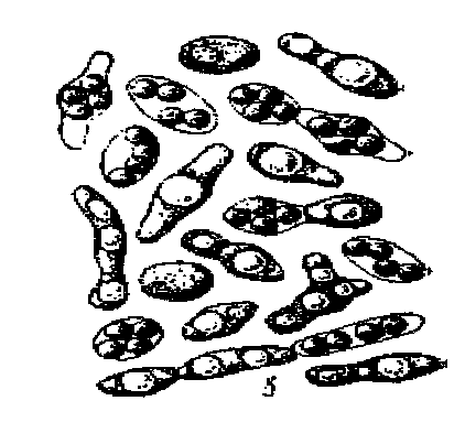
Morphologically yeast is diverse. They differ from each other with dimensions and form of cells. The sizes of yeast cells are dependent in the following limits; From 2.5 to 10 microns in the diameter and from 4 to 20 microns in length. The morphological variety of yeast forms is shown in Fig. 6.
but

b.
in

g.
 d.
d.

e.

j.

z.
Fig. 6.Forms of yeast cells: a - oval ovoid;
b - cylindrical; in - apiculant; lymonic; g - sweatshop;
d - triangular; e - sickle; Well - flaskoidal; s, and - Micepical
The shape and dimensions of yeast cells depend on the species, age, nutrient medium, the method of cultivation.
Depending on the type of yeast, it can grow vegetatively to multiply by killing (yeast of oval shape), binary division (characteristic of yeast cylindrical or rolling shape) or tick division. In addition to vegetative reproduction, yeast - ascomycetes can be broken by sex with the formation of Askospor.
From yeast belonging to the class of ascomettes, great importance have yeast-sugaromycetes of kindSaccharomyces. that widely used in food Industry. The main biochemical feature of these yeast is that they ferment sugars to form ethyl alcohol and carbon dioxide. Yeasts used in industry are calledcultural yeast. So, in bakery and in the production of alcohol, the horse yeast is used in the production ofSaccharomyces.cerevisiae.. Yeast speciesSaccharomyces.minor We found the use in the production of rye bread and kvass. Brewery uses lower yeastSaccharomyces.carlsbergensis. Yeast-sugaromycetes have an oval shape, vegetatively multiply by the kill, in adverse conditions multiply by sexual askospoda.
Some spore yeast arewild yeast . These yeasts are as well as cultivated, capable of alcohol fermentation, but in addition to alcohol form a lot of by-products (such as aldehydes, higher alcohols, ethers, etc.) and therefore worsen organoleptic product performance. These yeast are pests produced by various drinks (beer, wine, soft drinks), as well as pathogens of certain foods.
Yeast - deuteromycetes can be multiplied in a vegetative way. Some of these yeast (for example, yeast kindCandida.) Used in industry to obtain feed protein, organic acids, vitamins and other microbial synthesis products. Yeast speciesTorulopsis.kefir. Part of a symbiotic start-up - kefir fungus. Other representatives of imperfect (hydrodic) yeast are wild yeast and cause damage to many foods. Drainage-pests include man yeastPichia., Hansenula., Candida., Rhodotorula,Torula., Torulopsis., MyCoderma., Trichosporon. and others. Among the coophery yeast are foundfalse yeast that form pseudomycelions and grow on liquid substrates in the form of films.
Procedure for performing work
On the subject glass with a tube or pipette, a large drop of water is applied;
Select a small amount of mycelium from a test tube or Petri dishes, observing the Asepta rules
The mycelium is neatly placed in a drop applied to the slide and with the help of two needles lay it in water;
The drug is covered with covered glass and pressed slightly. Excess water is removed by filter paper.
Microscopes the drug "crushed drop" first with the lens X8, and then x40 in a darkened field of view (the condenser is omitted, the curtain of the iris-diaphragm is covered).
In the selection and microscopy of the drugs of mushrooms, the following recommendations take into account:
a) mushroom genus MUCOR. . Select the blackname-gray fluffy air mycelium. During microscopy draw attention to the gifs with the spores of the spores and the columns, which are formed when the sporangium is released;
b) genus kind Aspergillus. . Select a bit of fluffy mycelium with painted conidias, slightly deepening the needle into the nutrient medium. Pay attention to unfinished conidenos;
c) genus genus Penicillium. . When the selection is trying to take a young mycelium (on the border of the painted and white mycelium), deepening the needle on Wednesday. Pay attention to septic gifs with tassels.
d) mushroom genus Alternaria. . Take the fungne in black sites, deepening in her needles. Pay attention to septic myceliums, weakly developed conidiosses and large confidium, having a type of round or pointed multicellular formations resembling "lemon grenades".
When studying yeast A suspension of yeast is applied to the glassy glass, covered with coating glass, excess water remove with filter paper. Microscopic drug and lens x8 and x40.
Registration and analysis of research results
Briefly abstract theoretical material. They sketch microscopic patterns of the studied cultures of mushrooms and yeast, taking into account the morphological characteristics of each microorganism. Under each drawing sign the Latin name and increase in the drug. Describe the cultural properties of the studied mushrooms.
Answer control questions
How are microscopic mushrooms and yeast prepare prepare?
Describe the morphological and cultural properties of microscopic fungi.
What mushrooms are used in industry to produce organic acids, enzymes, antib0iotics and other valuable products?
Describe the morphological properties of yeast.
What are cultural yeast? In which food industry industries are they used?
Terms of completion of the task
Biology class
2. Maximum task execution time: 90 min
Practical work number 3: The schemes of the structure of the cells of bacteria, yeast, mushrooms.
Purpose of work: Examine the structure of the cellbacteria, yeast, mushrooms
Material support: instructive Cards for Practical Work, Textbook, Pencils
Exercise 1
Studies a textbook material. According to the results of the study:
Draw into the notebook the structure of the cells of bacteria, yeast and mushrooms and specify the distinctive features
Written to answer questions:
1. What form do bacteria cells have?
2. What are the sizes of bacteria?
3. What is the reproduction of bacteria, the reproduction rate?
4. What way, and in what conditions is the formation of a dispute with bacteria?
5. Are the bacteria to independent movement?
Take output by results.


Terms of completion of the task
1. Place (time) task execution
Biology class
2. Maximum task execution time: 90 min
Practical work on the topic №4 Work with regulatory and technical documentation: SanPine 2.3.6. 1079-01
Purpose of work: Examine sanitary requirements for the device and maintenance of catering
Material support: instructions for practical work, SanPine 2.3.6. 1079-01
Exercise 1
Studies a textbook material. SanPine 2.3.6. 1079-01. According to the results of the study:
1. Extract phrases: a plot where the catering company has been built, should be
The production premises include:
Warehouse premises are designed in ____________________ parts of the building.
Quality drinking water must match
Ventilation is used for air purification
Type.
All production facilities should be covered
Light.
Monthly cleaning of premises is called
2. Give the definition of the following concepts:
Disinfection is -
Deratization is -
Disinsection is -
3. Using educational material, Fill the table:
Vegetable shopMeat shop
Fish shop
Hot shop
Cold shop
Confectionary shop
Distribution
Terms of completion of the task
1. Place (time) task execution
Biology class
2. Maximum task execution time: 90 min
Practical work number 5 Work with regulatory technical documentation: SanPine 2.3.6. 1079-01
purpose of work : Examine sanitary requirements for equipment, inventory, dishes, containers. Transportation and storage of food products.
Material support : Instructive cards for practical work, SanPine 2.3.6. 1079-01
Exercise 1
Examine material textbook, SanPine 2.3.6. 1079-01. According to the results of the study:
1. Answer in writing to questions:
What does it belong to the kitchen utensil?
What is labeled dishes?
What does it belong to the dining room dishes?
What materials are allowed for the production of equipment and inventory
for catering enterprises?
What is the principled difference when washing the dining rooms and cutlery?
2. List the rules and requirements:
2.1. Sanitary rules Transportation of semi-finished products:
2.2. Sanitary rules for food storage:
3. Extract phrases:
Before the distribution, the quality of finished dishes should
When filing first dishes and hot drinks should have a temperature
_______ ° С, second dishes and side dishes ______ ° С, portion dishes
temperature ______ ° C, Cold dishes and drinks ______ ° C.
In therapeutic and preventive and children's institutions in the winter-spring period due to lack of vegetable dishes ___________________ It is required to enrich these some dishes.
For the quality of finished products and compliance with the rules of its holidays in catering enterprises are responsible ________________
Terms of completion of the task
1. Place (time) task execution
Biology class
2. Maximum task execution time: 90 min
Practical work number 6 The preparation of disinfecting solutions and their storage
Purpose: Examine the name of disinfectants, methods for preparing disinfecting solutions depending on the destination. Prepare a solution of a given concentration.
Material support : Instructive cards for practical work, SanPine 2.3.6. 1079-01, textbook
Exercise 1
Examine material literature, SanPine 2.3.6. 1079-01. According to the results of the study:
1. Answer questions:
What solutions relate to disinfectant?
What is the purpose of disinfecting solutions?
What drugs are used as disinfectors?
How to recognize what dishes were treated with disinfectors?
2. Examine the preparation and purpose of disinfectants. Fill out a table.
3. Prepare 1 liters 0.2% solution of chlorine B.4. Make a conclusion based on the results of work.
Terms of completion of the task
1. Place (time) task execution
Biology class
2. Maximum task execution time: 90 min
Criteria for assessments for practical work:
Rating "5" is set if
:
1. The correct independently determines the purpose of these works; Performs work in full compliance with the necessary sequence of conduct.
2. independently, rationally chooses and prepares the necessary equipment to perform work; Conducts data to work in terms of obtaining the most accurate results.
3. Competently, it logically describes the course of work, correctly formulates conclusions; Exactly and gently performs all records, tables, drawings, drawings, graphics, calculations.
4. Exhibits organizational and labor skills: supports the cleanliness of the workplace, the order on the table, economically spends the materials; Compliance with safety regulations when performing work.
Evaluation "4" is set if
:
1. Performs laboratory operation completely in accordance with the requirements for evaluating the results on "5", but allows for calculations, the measurements of two are three lack of one or one non-bug and one defects.
2. When executing the work, it allows inaccuracies in the description of the course of action; Makes incomplete conclusions when generalizing.
Rating "3" is set if
:
1.1 correctly performs work not less than 50%, but the volume of the completed part is such, which allows us to obtain true results and draw conclusions by the main, fundamental important tasks of work.
2. Selects equipment, material, starts working with the help of a teacher; Or during measurements, calculations, observations, makes an error, inaccurately formulates conclusions, generalizations.
3. Conducts work in irrational conditions, which leads to results with large errors; Or in the report admits a total of no more than two errors (in numbers records, measurement results, calculations, drawing up graphs, tables, schemes, etc.) that have no fundamental value for this work, but that influenced the result.
4. Allows a gross error during work: in explanation, in design, in compliance with the safety regulations, which the student fixes at the request of the teacher.
Rating "2" is set if
:
1. Does not determine the purpose of the work itself, cannot prepare the appropriate equipment without the help of the teacher; It does not fully work, and the volume of the executed part does not allow to make the right conclusions.
2. Allows two and coarsest errors during the work that cannot be corrected at the request of the teacher; Or produces measurements, calculations, observed incorrectly.
ATTACHMENT.
Attachment 1
Memo student
When performing work, the student must:
It is preliminary in detail with the theoretical material and to understand the microbiological patterns and the processes to be learned in practice.
Performing an experiment, comply with all precautions, a sequence of operations, carrying out the necessary observations.
Record the results of experience in notebook according to the scheme proposed in the work:
After the end of the work, put in order a workplace and pass it with a laboratory and a teacher.
In the cycle of laboratory and practically work on academic discipline "Basics of microbiology, sanitation and hygiene in food production", the device of microscopes, the main techniques used in microbiological studies are considered. With which the morphological, biochemical signs of bacteria are studied, sanitary and bacteriological assessment of objects ambient and food products. Rules for the preparation of disinfecting solutions and rules for conducting equipment for equipment, inventory, dishes, containers. Prevention of production injuries, assistance.
Download:
Preview:
Laboratory and practical work
by discipline
Basics of microbiology, sanitation and hygiene in food production
Methodical instructions for students
Profession ____ 260807.01 Cook, confectioner __________
Tarasovo 2015.
Compiler: Repenko Z.V., teacher
Introduction | |
| |
The simplest microbiological research | |
| |
| |
| |
Differentiated test | |
Bibliography |
Introduction
Educational discipline OP.1Basics of microbiology, sanitation and hygiene in food production enters the structure of the general professional cycle.
The number of hours to master the program discipline program:
maximum Training Training Load51 hours, including:
mandatory audit learning load study36 hours;
independent work studying12 hours;
consultations - 3 hours.
In the cycle of laboratory and practically work, it is necessary to consider the device of microscopes, the main techniques used in microbiological studies. With the help of which the morphological, biochemical signs of bacteria are studied, a sanitary and bacteriological assessment of environmental objects and food products is carried out. Rules P.mitigation of disinfecting solutions and sanitary processing of equipment, inventory, dishes, containers. Prevention of production injuries, assistance.
Conducted laboratory and practical work will allow students to consolidate the knowledge gained in the class of theoretical course, and master the skills of microbiological research and sanitary processing of equipment, inventory, dishes.
The goals and objectives of the educational discipline are the requirements for the results of the development of discipline:
As a result of the development of educational disciplineshould be able to :
comply with personal hygiene rules and sanitary requirements for cooking; produce sanitary processing of equipment and inventory;
prepare solutions of disinfecting and detergents;
perform the simplest microbiological research and evaluate the results obtained.
Laboratory work №1 (a)
The simplest microbiological research
Purpose: study of the rules of work in microbiological laboratories and general rules Working with a microscope.
Duration: 1 hour
Equipment: microscopes.
Classes:
1. Organization of microbiological laboratories and rules of work in them.
2. Microscopes and microscopic equipment.
Examine the rules for working with a microscope.
Rules of work in microbiological laboratories
When working in the microbiological laboratory, the learner is obliged to strictly follow the rules of the internal regulation.
1. Everyone must work in coats, caps and replaceable shoes
2. In the laboratory it is forbidden to smoke and eat food.
3. The workplace must be contained in the sample order.
4. When accidentally hit the contagious material on the table, gender, etc. This place must be carefully treated with a disinfectant solution.
5. Storage, observation of cultures of microorganisms and their destruction should be made according to the special instructions.
6. At the end of the work, the hands should be carefully flushed, and if necessary, treat a disinfectant solution.
Methodical instructions:
General rules for working with a microscope. Work with any microscope consists of the correct installation of the illumination of the field of view and the drug and its microscopy with different lenses. Lighting can be natural (daily) or artificial, for which special light sources use. The place for the microscope is chosen further from direct sunlight. Work on the table with a dark surface less tires the eyes. It is better to look into the eyepiece with the left eye without closing the right.
Tolerate a microscope, holding one hand for a tripod, the other for the base of the microscope. It is necessary to protect the microscope from the jokes, contact with potent acids, alkalis. It is not recommended to remove the eyepiece from the pipe so as not to contaminate the pipe and lenses. During work, it is desirable to protect the microscope from breathing, as the condensation of vapors leads to it.
Lenses should always be clean. The microscope should be stored in the case. It is impossible to touch the optical surfaces with the fingers.
In microscopy of drugs, it is strictly followed by a certain order in operation:
1) Cooked and painted strokes to put on the subject table (to strengthen the clamps necessarily);
2) establish the lighting so that a light ring of the diaphragm appears in the field of view;
3) Rotate the revolver to the required lens (up to click);
3) carefully omit the microscope tube before the objects appear;
4) Conduct the final focusing of the drug with a micrometric screw, rotating it within only one turn. It is impossible to contact the lens with the drug, as this can entail the breakdown of the drug or frontal lens.
At the end of the work, the microscope is rubbed and removed into the case, the slide glasses are washed and dried.
Control questions:
- Why so many rules of behavior in microbiological laboratories?
- How is a microscope stored?
- Tell the microscope transfer rules.
Laboratory work number 1 (b)
The simplest microbiological research
Purpose: Consideration of preparation options.
Duration: 1 hour
Equipment: Microscopes,
Classes program
1. Preparation of drugs for the study of living cells.
2. Preparation of drugs fixed.
Task for performing laboratory work:
Examine the technique of preparation of drugs.
Methods of preparation of the drug:
The test tube with a culture is kept in the left hand almost in a horizontal position near the burner. The bacteriological needle from the tube is burned in the flame, a small amount of microbial mass takes. Before taking a culture with his right hand, take a cotton jam from the test tube, clamping it between the little finger and the palm, and the edges of the tube burn on the burner flame. The needle holds in the right hand big, index and middle fingers.
If the culture is taken from a liquid medium, should not be very tilted with a test tube so as not to moisten its edges and a plug. For the capture of culture it is better to use the loop. After taking the culture of the edge of the test tube and the plug burned in the flame and closed the test tube.
- The study of living cells of microorganisms by the methods of "crushed" and "hanging" drops. Both methods are used to identify the mobility of cells of microorganisms, observing the reproduction, formation and germination of the dispute, establishing the reaction of microorganisms to chemical compounds and physical factors of the effect, the study of cell size, the nature of their location and the determination of the spare substances of the cell.
Microscopic drugs, slightly dimming field of view; The condenser is slightly lowered, the flow of light is regulated by a concave mirror. Initially, use a small increase - lens 8x, after they detect the edge of the drops, set the lens 40x.
The method of "crushed" drop. A drop of tap water is applied to the clean glass. It makes culture and mix with water. Cover a drop with coating glass so that air bubbles are not formed under it. The glass wand presses the coating glass to the subject and remove the excess water with filter paper, bringing it to the edges of the coating glass.
Method "hanging" drops. Apply for long-term observations of cells of microorganisms. The sterile covered glass is caused by a needle, a spinning suspension of microorganisms grown in a liquid nutrient medium or prepared for this purpose in a physiological solution (0.5% NaCl solution). The covering glass is turned over and placed on a sterile subject with a hole in the middle so that the drop freely hung over the hole. For tightness, the edges of the hole are lubricated with vaseline.
- Fixed microorganisms.Fixed preparations are often prepared in microbiology. They are considered under the microscope painted. Under fixation implies such processing of a living object, which makes it possible to quickly interrupt the course of life processes in it, retaining a thin structure. As a result of fixing, the cells are firmly attached to the glass and better score. Fixation is necessary in case of working with pathogenic microorganisms for safety.
Capture a smear. A drop of tap water is applied to a clean skimming glass. The calcined bacteriological needle of the test tube with culture take a small amount of microbial mass and brought to a drop. The drop thoroughly smear the loop on the glass on the square of approximately 4 cm2 . The suspension of normal thickness is smeared with a thin layer on the glass, then the smear is dried in air at room temperature or weak heating, holding the drug high above the burner flame. Strong heating of the drug during drying is not recommended, since proteins coagulate, distorting the structure and shape of the cells. Dried preparation fixed.
Fixing the smear. Perform over the flame of the burner in the study of the shape of the cells. In the first case, the drug three or four times is slowly conducted by the bottom side above the burner tribe.
Coloring the drug. A few drops of dye are applied on the smear. Depending on the type of dye and the purpose of the study, the duration of staining changes from 1 to 5 minutes, in some cases up to 3 minutes and longer. At the end of the staining, the drug was washed with water, water is removed by filter paper, dried in air and microscopy.
There are simple and differentiated coloring methods. With simple coloring, a single dye is used, such as methylene blue, fuchsin, violet gential in alkaline or carbolic solutions. The whole cell is scratched. With differentiated coloring, individual cell structures are painted with different dyes. These are coloring methods by gram, painting dispute.
Control questions:
- Tell the principle of preparation of the drug by the method of "crushed" drops.
- Tell the principle of preparation of the drug by the "hanging" method.
- How is the fixation of the smear?
- How is the painting of the drug?
Laboratory work number 2.
The simplest microbiological research
Purpose: study of various forms of microorganisms
Duration: 2 hour
Equipment: microscopes.
Classes program
1. Microscopation of prepared drugs.
2. Filling reports.
Task for performing laboratory work:
Examine the forms of bacteria, mushrooms, yeast.
Method of execution:
- Examine the shape of mushrooms genus Penicilium.
Caution with the help of two vacuer needles, a piece of mycelium is removed from the medium and placed in a drop of water on the slide. The top glass (the crushed drop method) is placed on top.
A glass wand or prepar needle is slightly pressed on the center of the coating glass. Excess water is removed by filter paper.
The drug is viewed first with a small magnification, paying the main attention to the edges, since they are usually clearly visible to the brush of the conidiones. When the appropriate site is found, transition from the lens 8x to the lens 40x and the brushes are considered in detail.
- Examine the shape of bakery yeast.
Multiply by killing. When you kill on the maternal cell there is a small convexity - "kidney" is a subsidiary in which one core passes, the cell increases in size and separated. If conditions for such reproduction are favorable (sufficient sugar, suitable temperature, aeration), the process is very fast. In some representatives of the kind of cells, after kinding, do not have time to disconnect and the pseudomitiates arise (false mycelium).
A small piece of yeast mass a few hours before classes are placed in warm creamy water and put in a warm place. Middle liquid is formed. It is applied to the slide glass, dried in air. Cells are clearly visible with smaller increases.
Two races are usually present in bakery yeast: one is represented by rounded-ellipseed cells, quickly disconnecting during the scouring; Another - elongated-cylindrical, forming branching bushes (pseudomytellius). Many cells are visible kidneys. In the fine-grained content of living yeast, large transparent vacuoles occupy the central position are well noticeable.
- Examine the microflora of the oral cavity.
Using toothpicks, apply to the slide tooth flare. Conduct fixation, handle the coloring substance (solution of fuchsin), rinse, remove excess water with filter paper, dry in air and microscopy.
application
Fig. 1. Bacteria shape:
character: A - micrococci; b-diplococci; E - Tetracockers; g-streptococci; d - staphylococci; E - Sarcin; chopped; Well - not forming dispute; s, and, k - spore-forming (z - bacillalar, and - clostridial, to - plexldial types of spioning); Sorry: L - Vibrini;m - spirilla; n - spirocheti
Fig. 2. Microscopic mushrooms:
but - Misogne; b - Aspergillus; in- Penicillium; a - fusarium; d - trichoderma;
E - Alternarla; Well yeast ticking; W - Delyads
Fig. 3. Yeast Saccharomyces Cerevisiae in the recipigation stage
Control questions:
- List the factors affecting the development of microbes
- What is the optimal temperature of the development of mold fungi and yeast?
- Describe the shape of bakery yeast.
- What are the forms of the bacteria of the oral cavity?
Laboratory work number 3.
Preparation and analysis of disinfecting solutions
Duration: 2 hour
Equipment: Chlorine lime (deo - chlorine), microscopes.
Classes program
1. Preparation of disinfecting solutions of different concentrations.
2. Studying washes from equipment.
Task for performing laboratory work:
Study the effect of disinfecting solutions for microorganisms
Method of execution:
1) In catering establishments, disinfection is carried out with a prophylactic goal to prevent the possibility of infection with microbes of food and finished food. For disinfection, physical and chemical methods use.
When choosing these funds for public catering enterprises, you should pay attention to:
Registration certificates indicating the possibility of using disinfectants at catering;
Certificate of conformity - a document confirming the compliance of this disinfectant to the requirements of the standard;
Instructions for the use of disinfectants.
Chlorine lime (inorganic substance), solutions of different concentrations of which are used for disinfection of premises of catering, equipment, equipment, dishes. At the same time, vegetative and dispute shapes of microbes are destroyed. Usually a 10% clarified solution of chlorine lime is prepared, dissolving 1 kg of dry chlorine lime in 10 liters of water and insisting it within 24 hours in a glass dish in a dark place. This solution is stored for 5 days and is used to obtain solutions of a lower concentration by diluted with water;
Method for preparing disinfectants
p / P. | Name | Concentration,% | Purpose | Cooking method |
Bleaching powder | 10 (source) | Processing containers for food waste | 1 kg of chlorine lime on 10 liters of water, uphold 24 hours, merge with precipitation |
|
Processing shells, washbasins, toilets | 5 liters of starting solution dissolve in 10 liters of water |
|||
Disinfection of equipment and inventory | 2 liters of the starting solution dissolve in 10 liters of water |
|||
1 (working) | Room processing (floors, walls, doors, etc.) | 1 liter of the initial solution to dissolve in 10 liters of water |
||
Equipment processing | 0.5 liter of the initial solution to dissolve in 10 liters of water |
|||
0.2 liter of starting solution dissolve in 10 liters of water |
||||
Chloramine B. | Disinfection of dining room dishes, hands | 20 g (1 tbsp. Spoon) dissolve in 10 liters of water |
||
Disinfection of premises, equipment | 50 g (2.5 tbsp. Spoons) dissolve in 10 liters of water |
|||
Hypochlorite calcium | Disinfection of dining room dishes | 10 g (1ч. Spoon) dissolve in 10 liters of water |
2) study the effect of disinfecting solutions for microorganisms
With the help of a cotton sticks to apply a washed glass on the slide glass. Conduct fixation, handle the coloring substance (solution of fuchsin), rinse, remove excess water with filter paper, dry in air and microscopy. Process equipment with a disinfectant solution, prepare a re-preparation and microscopy.
Control questions:
- What forms of bacteria are on the surface of the equipment?
- How microorganisms react todisinfecting solutions?
- What is the concentration of the initial solution?
Laboratory work number 4.
Sanitary processing of equipment, dishes, inventory
Purpose: The formation of skills to prepare disinfecting solutions for equipment processing, inventory, dishes
Duration: 4 hours
Equipment: Disinfecting solution, technological equipment of the culinary and confectionery shop.
Classes program
1. Preparation of disinfecting solutions of the required concentration.
2. Study of equipment processing rules, inventory, dishes.
Task for performing laboratory work:
Examine the rules for processing equipment, inventory, dishes disinfectant solutions
Method of execution:
- Examine sanitary-epidemiological requirements for equipment, inventory, dishes.
- Process equipment, equipment, dishesdisinfecting solutions of the necessary concentration.
- Based on the previously obtained knowledge and skills, draw conclusions about the need for timely sanitary processing of equipment, inventory, dishes.
Control questions:
- How to wash and disinfect mechanical equipment, including with removable work units?
- What sanitary requirements are presented to the device and content of production tables?
- What sanitary requirements are presented to the content of thermal equipment?
- What is the value of the marking of cutting boards, knives?
- What is the sequence of washing the dining room dishes manually in the washing baths?
Practical lesson number 1
Prevention of production injuries, assistance
purpose the formation of skills to avoid production injuries and provide first prefigure help
Classes duration - 4 hours
Tasks:
Send the ability to prevent production injuries.
Develop the ability to first-bear the victim
Equipment: Typical Safety Instructions, Additional Theoretical Material for First Prompt Assistance to victims (with illustrations)
The task: examine the proposed theoretical material and perform a practical building: conduct safety instructions; Render the first help victim.
Labor protection and safety
1. Legislation on labor protection and safety
The protection of workers' health, ensuring safe working conditions, the elimination of occupational diseases and industrial injuries is one of the main worries of our state.
In accordance with the Constitution of Russia, citizens are ensured equality in the field of labor, regardless of nationality and gender. A woman is provided with equal rights with a man for labor, his payment, rest and social security.
Protection of labor rights of citizens is carried out state organizations and professional unions. In the basics of the country's legislation, great attention is paid to creating favorable working conditions for human life and human health. It includes a complex of legal, technical and sanitary and hygienic events.
Occupational safety activities are developed on the basis of the country's Constitution, and their implementation is assigned to the administration of enterprises and organizations. The organization is obliged to introduce modern means of protection, warning production injuries and ensuring sanitary and hygienic conditions that prevent occupational diseases.
Labor protection in Russia is a wide range of legal, sanitary and hygienic, technical and organizational measures aimed at creating healthy, safe and high-performance working conditions at catering establishments.
Safety is one of the main tasks of labor protection, which includes a complex of technical and organizational measures aimed at creating and implementing safe techniques, safe production processes, automatic communication tools - and alarm, fender and safety devices, as well as personal protective equipment, Preventing the possibility of industrial injuries.
At each enterprise, the relationship between workers and employees with the administration is negotiated in the form of a collective agreement, which lies with the local trade union committee on behalf of workers and employees with the administration of the enterprise. The conclusion of the collective agreement is preceded by the discussion and approval of its project at the meeting of workers and employees. This contract applies to all workers and employees of the enterprise, regardless of whether it consists by a member of the trade union.
The collective agreement contains the basic provisions on labor and wages established for this enterprise, in accordance with current legislation, as well as provisions in the field of working time, resting time, wage and material incentives, labor protection, developed by the enterprise administration and the trade union team within the limits of rights provided to him.
Legislation, protecting the established duration of the working day (40 hours), a week as a rule, does not allow overtime. Such work is allowed in exceptional cases, but even in the presence of legal grounds for overtime, the enterprise administration is not in the right to carry them out without permission of the trade union committee.
Labor legislation shows an exceptional care for the younger generation and provides for the most favorable conditions for labor, recreation and teenage training.
Reception is allowed, starting from 16 years old, with six hours working days, while saving payment for full-time as adult employees of the corresponding category. It is forbidden to use the work of adolescents in the night and overtime. Teens are not allowed to work with harmful and heavy production conditions.
All workers and employees have established an annual paid leave by a duration of at least 24 working days. Women are provided with many other benefits in accordance with the current legislation.
The administration of public catering is required to provide issuance, storage, washing and repair of workwear, specialobuvi and other personal protective equipment. Control over compliance with the implementation of labor protection laws, safety and industrial safety laws is carried out by the State Inspection Bodies for Work and Professional Unions. Control over compliance with enterprises of sanitary and hygienic working conditions - the State Sanitary and Epidemiological Service, and for compliance with fire safety enterprises - the State Fireman Supervision.
The trade union committee provides also control over the work of the catering company and fulfill the administration of labor legislation, rules and safety standards and industrial sanitation. With the failure to fulfill obligations under the collective agreement, non-compliance with the norms and rules for labor protection and safety regulations, the trade union committee has the right to raise the question of punishment or removal from the position of executives of the enterprise.
2. Organization of work on labor protection
Work on labor protection in enterprises should be organized in accordance with the Regulation on the organization of work on labor protection, developed taking into account the current sectoral provision on the organization of work on labor protection and approved by the head of the enterprise.
The situation should indicate that general leadership and responsibility for the organization and work on labor protection as a whole on the enterprise is assigned to his head (owner), and in the structural divisions of the enterprise - on their leaders.
At the enterprise the situation must be set to the order:
Organization and frequency of training for labor safety workers;
Conducting and frequency of integration of labor safety;
Work on fire safety;
Carrying out work of increased danger with the issuance of the admission outfit;
Carrying out loading and unloading;
Maintenance of equipment;
Consolidation of equipment for people responsible for its proper and safe operation when using it;
Ensuring and issuing workwear workers and personal protective equipment;
Control over the observance of rules and labor protection standards on the enterprise as a whole and its structural divisions.
Practical work on labor protection is carried out by a special service, a labor protection engineer or a person, which is entrusted to the order for the enterprise, this work subordinates directly to the company's head.
Training of labor safety workers should be carried out at all public catering enterprises, regardless of the nature and degree of danger of production, as well as independently of the forms of ownership.
Instructing and training for safe techniques and methods of work is carried out for all working and engineering and technical workers in all areas, regardless of the experience, qualifications and experience of the working, as well as for persons who arrived at the enterprise for passage industrial practice.
In catering factory, labor safety instructions in the nature and time of the presentation are divided into the introductory, primary in the workplace, repeated, unscheduled and target.
Induction training. The introductory intention on occupational safety is carried out with all the newly adopted work, regardless of their education, work experience in this profession or position, with temporary employees, commaed, students and students who arrived in production practices.
The introductory instruction is carried out according to the program approved by the company's head. This briefing should be carried out by the head of the enterprise or an employee, whom the order of the head of the enterprise is entrusted with practical work on labor protection and technology
security.
When entering an introductory safety instruction, the enterprise administration is obliged to familiarize the employee:
With the main provisions of labor legislation;
With the rules of the internal labor regulation;
With the basic requirements of electrical safety;
With the procedure for drawing an accident on the accident associated with
production;
With the procedure for providing first aid victims of electric current and with other accidents;
With the general requirements for the organization and maintenance of workers
places;
With the requirements of personal hygiene and industrial sanitation, the appointment and use of Sanitarywood, San assecobuvi and safety devices.
On the conduct of introductory instruction makes entry in the logging log on the registration log with a mandatory signature of the instructable and instructors, as well as in the work document. Along with the magazine, a personal learning card can be used.
Primary briefing. Primary briefing in the workplace should be held all the newly incoming workers and students sent to businesses for the passage of production practices, as well as workers translated from one job to another or from servicing one type of equipment to another.
Without briefing in the workplace, no employee should be allowed to work.
Instructions in the workplace should be carried out by the heads of those structural units, in direct subordination of which will be instructed employees.
In small enterprises that do not have structural divisions, the instruction is assigned to the head of the enterprise.
When carrying out an instruction on safety in the workplace, the employee must be familiar in detail:
With the equipment of the equipment on which Ra Batnik will have to work and which it will serve;
With all dangerous places from the car, with safety fences, adaptations and means of individual protection, with their appointment and rules of use;
With the right and secure service organization *! working place;
With the procedure for preparing for work (checking the health, grounding, tool, inventory, etc.);
With the safe and correct techniques and the consequences of the application of incorrect work techniques;
With the safety instructions for the safety of the equipment;
With the order of safe movement in the territory of the enterprise.
The instruction must be accompanied by a show at the site of the right techniques for working with the repetition of employees of these techniques. The instructrive must be convinced of a clear knowledge and understanding of the safety regulations by each employee.
Repeated briefing. Re-briefing at the workplace should pass all employees, regardless of the qualifications, education and experience of the work. It is held with the goal of better assimilation, deepening and consolidating knowledge on safe techniques and methods of labor.
If, as a result of the inspection, unsatisfactory knowledge of the safety instructions will be revealed, instructing is obliged to give an employee all the necessary explanations and directly in the workplace to show how it is necessary to work properly safe methods and require the strict implementation of all the requirements of safety instructions. The instruction should be supported by a detailed analysis of examples from the practice of the enterprise.
Unscheduled briefing. An unscheduled instruction is carried out:
With the introduction of new or recycled standards, rules, labor protection instructions, as well as changes to them;
When changing the technological process, replacing or upgrading equipment, devices and tools, raw materials, materials and other factors affecting the safety of labor;
In violation by the worker, the safety requirements of labor, which can lead or led to injury, accident, or fire, poisoning;
At the request of supervision bodies;
For breaks in the work - for the work to which additional (elevated) labor safety requirements are presented - more than 30 calendar days, and for other works - 60 days.
Target briefing. Target briefing is carried out in the fulfillment of one-time work, unrelated with direct responsibilities in the specialty, elimination of the consequences of accidents, natural disasters and disasters, the production of work on which the outfit, permission and other documents are made. All types of briefing are drawn up in a special logging log of the installed form. Pages of the magazine must be numbered, laid and fasten the seal.
In accordance with the requirements of the health authorities, each employee of public catering passes periodic medical examinations.
The frequency of medical examinations that the employee must pass during operation are established in accordance with the requirement of health authorities. The employee of catering enterprises is obliged to have a personal medical record in which the results of medical examinations are made.
Norms are installed on catering establishments for raising and moved gravity:
for women:
When alternating with another work (up to 2 times per hour) weighing more than 10 kg and constantly during the work shift - weighing no more than 7 kg;
The magnitude of the mass of the moved cargo or the lifting per shift when lifting the working surface should not exceed 5 tons. From the floor or level below the working surface - 2 tons.
for men:
Constantly during the work shift weighing no more than 30 kg (a load-loader - no more than 50 kg);
The magnitude of the mass of the cargo being moved or lifted for the shift (in all the works besides loading and unloading) when the working surface is lifted, it should not exceed 12 tons, from the floor or level below the working surface - 5 tons.
for teens from 16 to 18 years old:
If this work takes no more than 1/3 of working time - weigh no more than 16 kg;
With constant transfer of gravity - weighing no more than 4 kg. The distance between workers carrying loads must not be
less than 3 meters.
3. Production injury
An accident or injury is called an incident in which as a result of the sudden impact (mechanical, chemical, thermal) external environment, there has been damage to human organs or violation of their normal life activity.
The production is considered to be an injury received by a worker or serving when performing its employment duties, when performing an action in the interests of production or way to work and from work on transport submitted by the Organization.
At the enterprise of catering cases of injury, mainly with the process of cooking. Injuries occurring as a result of violation of safety regulations and work discipline.
All cases of industrial injuries are subject to consideration and accounting. Acute poisoning, thermal blows, frostbite do not relate to industrial injuries, but are taken into account as accidents. All industrial accidents, no matter when they have occurred, are subject to careful investigation and adopting appropriate measures to their unagnation.
On the accident in production, the injured or an eyewitness is obliged to inform the director of the enterprise or the responsible for production. The victim is assisted, and if necessary, cause a doctor. Investigation are subject to all accidents on production, which cause loss of disability for a period of one day or more. The head of the enterprise in conjunction with the public inspector for labor protection and the responsible worker for labor protection at production for 3 days is jointly investigated and the Act shaped N-1 in four copies is made.
Investigation are also subject to minor accidents without disability as the causes of them causing them, they can lead to more severe production injuries.
The enterprise administration is obliged to analyze all accidents, while developing specific measures to eliminate and control their implementation.
4. Fire safety
In our country, there is a special body for the organization of fire protection - the State Fireman Supervision. Its task includes the development and implementation of measures to eliminate the causes of fires.
Fires, as a rule, arise as a result of violations and ignorance of fire safety rules. Therefore, regular briefing on fire safety measures is important to prevent fires.
Production and warehouses contain clean and order. After the end of the work, carefully inspect: electrical equipment (except refrigerators) must be turned off, gas equipment is turned off by a crane on the domestic gas pipeline, the workshops are carefully removed.
Use only good switches, rosettes, forks, cartridges and other electric machines.
Not to leave unattended equipment and electrical appliances. Upon completion of work, turn off the electrical lighting (except for emergency).
Smoking only in specially designated and equipped places.
Passages, exits, corridors, stairs, tambura keep clean, without cluttering the package and other objects.
The company must have permanent primary fire extinguishing facilities.
In catering establishments, the main causes of the fire can be: careless handling of fire, the unsatisfactory technical condition of electrical equipment, a malfunction of thermal equipment and drying on them workwear, etc.
The basic principles of fire extinguishing are - cooling a combustible substance below the temperature of its ignition and isolation of it from access of oxygen of air or other oxidizing agent that supports combustion. Most of the extinguishing means applied to the fire extinguishing means of combustion comprehensively - stops the access of the oxidant and prevents the transmission of heat from the flame to be combustible, at the same time enhance heat transfer into the environment.
The main means of fire extinguishing include - water, water vapor, air-mechanical and chemical foams, inert and carbon dioxide, powdered dry compounds from the coarse soda, sand and various bedspreads from asbestos, tarpaulin and other faded materials.
Each public catering worker must comply with the existing fire safety rules. When the fire is detected or signs of burning (smell of smoke, the smell of gary, temperature rise, etc.) is necessary:
Stop working and disable using the "Stop" button (switch, switch, crane, etc.) used equipment and electrical appliances;
Immediately inform about this by phone in the fire protection;
If possible, measures for the evacuation of people, fire extinguishing and the safety of material values.
5. Major safety activities in production
Currently, it is difficult to imagine any enterprise without the use of electrical energy. Moreover, public catering enterprises where various types of technological electrical equipment are used for cooking and eating.
The widespread use of them leads to the need for just as wide training of service workers with the rules of safe operation of electrical equipment, since the violation of these rules leads to damage to equipment, fires and death of people.
When a person is in the sphere of action of an intense electromagnetic field or directly contacts the electrical current under voltage, an electric current passes by its body. As a result of the operation of the electric current to the body, an electrician may occur, that is, more or less significant violations of functions.
The nature and intensity of disorders in the body caused by an electric current is mainly determined by the type and value of the current with a duration of its action and a number of other factors.
The defeat of the human body is more dependent on the magnitude of the current passing through the vital organs of a person - the brain, the central nervous system, the heart, the boding controls and from the individual characteristics of the victim.
All electric shocks are divided into two types - electrical injuries and electrical shocks. Electric strikes are most dangerous, since they cause violation of physical processes in the human body.
In order to avoid the defeat of working personnel by electric shock at catering establishments, individual and general protective equipment are used.
Individual protective equipment includes dielectric gloves, rugs, galoshes and insulating stands. Recommended when working with electrical equipment to have dry hands, clothing and shoes.
The total means of protection against damage include protective grounding, reassembly, and automatic shutdown of equipment.
Equipment operating on gas fuel is an increased danger, since the gases of poisonous and inhalation can cause poisoning.
In addition, gas in a certain ratio with air forms an explosive mixture, which explodes from the slightest spark.
That's why the main security activities are made by safety issues with gas equipment.
Fire safety of enterprises should be ensured by fire prevention and fire protection systems, including organizational and technical measures based on existing legislation on labor protection.
Grounding (rewarding) are subject to:
The buildings of all electrical apparatus, machinery and equipment installed at catering;
Drives of electrical apparatuses;
Frames of switchboards and control panels, cabinets, if electrical equipment is installed, voltage above 42 V AC;
Metal housings of mobile and portable electrical receivers;
Electrical equipment installed on moving parts of machines and mechanisms.
That is why the health of the protective ground (reinforcement) or the protection system is of great importance to prevent electrical exchanging in catering enterprises. However, you need to remember and do not forget when cleaning the room or electrical equipment, that water and wet rag are a good electrical current conductor. Therefore, it is strictly forbidden to put wet overalls, metal items on electrical equipment and supplying devices.
6. Typical instruction for labor protection for cook
1. General safety requirements
In order to avoid an accident at work, the cook is obliged to carry out instructions for labor protection.
Men and women are allowed to work as a cook, not under the age of 18 years old, trained in the specialty.
At the workplace, the cook receives primary safety instructions and passes an internship with the rules of operation of technological equipment attached to it.
When operating a gas-grade equipment, a cook before appointment on independent work It is obliged to undergo training in safe methods and acceptance of work in gas economy and pass exams in the prescribed manner.
During operation, the cook should take:
Inspection of open surface surfaces for diseases - daily;
Labor safety training on existing equipment - every 2 years;
Re-checking knowledge of safe working methods and techniques for work in gas economy - annually;
Verification of electrical safety knowledge - annually;
Check sanitary and hygienic knowledge - annually;
Periodic medical examination;
Re-instructing on the safety of labor at the workplace, the cook should receive once every 3 months;
Each cook should be provided with sanitary clothing, shoes, sanitary entrepreneurs and personal protective equipment.
2. Security requirements before work
The cook is obliged while working to wear sanitary clothes relying to him: the hair is removed under the headdress, the sleeves of the clothes are rented to the elbow or fastened by the hand. It is not recommended to challenge the sledgewood needles and keep in pockets pins, glass and other beating and sharp objects.
Before starting work, the cook is obliged to put in order its workplace for safe work and check:
Serviceability and idle equipment;
The presence and health of fences;
The presence and health of grounding;
The serviceability of other equipment used;
Make sure that the switches of the electric stove and the roast cabinet are in the zero position;
Control and work of local exhaust ventilation, air scenario.
If any problems or malfunctions are found in the equipment, the cook is obliged to immediately declare the workshop or the administration of the enterprise and before eliminating them to work do not start.
3. Safety requirements while working
To prevent the adverse effect of infrared radiation on the body, the cook must:
Fill out the work surface of the electric stoves as much as possible, turn off the electrolycit sections in a timely manner or switch them to a smaller power;
Prevent the connection on the maximum and middle power without loading;
Do not allow liquid to enter the heated burners of the plate, pumped up the dishes to fill no more than 80% of the volume;
Do not use porch boilers, saucepans and other kitchenware, having deformed bottom or edges, fragile knobs or without them;
To shoot from the plate with hot food without jerks, observing caution, together using dry towels or mittens, the boiler lid should be removed.
Control pressure and temperature in thermal vehicles within the limits specified in the operating instructions.
Monitor the presence of thrust in the combustion chamber of gas-wide equipment and testimony of pressure gauges during operation of a pressure-operating equipment.
4. Safety requirements in emergency situations.
When malfunctions are detected when working with mechanical, steam, electrical and gas equipment, as well as when the safety valve is triggered, the water, the leakage of water must be immediately disabled, inform the head of production or the enterprise administration.
Before eliminating observed problems, it is not recommended to start work.
Without permission, the administration is not allowed to produce any repair of equipment or troubleshoot.
5. Security requirements at the end of work.
Before disconnecting from the electrical network, you first need to turn off all electrical equipment with the exception of duty lighting and equipment operating in automatic mode.
After disconnecting gas-wide installations, remove the caches from cork cranes.
When conducting sanitary treatment, do not cool the heated surface of the plates, frying pan and other thermal equipment with water.
Provision of first prefigure
Whatever misfortune happens - in any case, first aid should be started with the restoration of cardiac activity and breathing. Then proceed to a temporary stop of bleeding. After that, you can proceed to the imposition of fixing dressings and transport tires.
As practice shows, it is such an order of actions that will help preserve the life of the victim prior to the arrival of the medical personnel.
First Aid Procedure:
1. If there is no consciousness and there is no pulse on the carotid artery - to proceed to resuscitation
2. If there is no consciousness, but there is a pulse on the carotid artery, - turn on the stomach and clean the oral cavity
3. With arterial bleeding - impose a harness
4. If there are wounds - impose bandages
5. If there are signs of fractures of the bones of the limbs - to impose transport tires.
SUDDEN DEATH Signs of sudden death (when every lost second can become fatal) 1. Lack of consciousness. 2. There is no reaction of pupils into light. 3. No pulse at the carotid artery. Signs of biological death (when resuscitation is meaningless) 1. Drying the cornea of \u200b\u200bthe eye (the appearance of a "selegant" shine). 2. The deformation of the pupil with the careful compression of the eyeball fingers. 3. The appearance of body spots. If there is no consciousness and there is no pulse on the carotid artery 1. Ensure the absence of a pulse on the carotid artery (Fig. 1). It is impossible! Saying time to determine the signs of breathing. 2. Release the chest from clothes and unzip the belt belt (Fig. 2). It is impossible! Apply a blow to the chest and carry out an indirect heart massage without freeing the chest and not unbuttoning the belt belt. 3. Cover the swordeephoid process with two fingers. It is impossible! Apply a blow to a mildo-shaped process or in the region of the clavicle (Fig. 3). 4. Apply a punch in the chest. Check the pulse. If there is no pulse - go to the next position 5 (Figure 4). It is impossible! Apply shocks in the presence of a pulse on the carotid artery. 5. Start indirect heart massage. Pressing 50-80 times per minute. The depth of priming the chest should be at least 3-4 cm. It is impossible! Press the palm on the chest so that the thumb is directed at the rescuer (Fig. 5).
7. With the narrowing of pupils, but the absence of heartbeat resuscitation must be carried out before the arrival of the medical staff. Arterial bleeding Signs of arterial bleeding 1. Alay blood from the wound beats a fountaining jet. Signs of venous bleeding 1. Blood passively flows from the wound. 2. Very dark blood color. In cases of arterial bleeding 1. Press the artery fingers or fist at the specified points. (Places of pressing large blood vessels.) Before applying a harness, the damaged limb should be left in a raised position. On the limbs, the point of pressed artery should be above the place of bleeding. On the neck and head - below the wound or in the wound (Fig. 7). It is impossible! Lose time to release the limbs from clothes.
Wrap the loop-closure around the harness. Press the loop and start the free end of the harness. Attach a note on the overlay time of the harness under the gum of the loop (Fig. 8). Harness to the limb can be imposed no more than 1 hour. Harness on the neck impose without controlling the pulse and leave until the doctor arrives. To seal the wounds use a clean napkin or multilayer fabric (Bandage packaging). In cases of scoring and edema of the limb (with improper imposition of a harness), it is necessary to immediately apply harness. The harness on the hip is superimposed through a smooth subject (bandage) with the control of the pulse on the popliteate yam. Wounding limbs How to impose bandages on wounds 1. To cover the wound by any pure napkin, fully glusted the edge of the wound. Forbid! Rinse with water with water (Fig. 9). 2. Bake the napkin or glue it with adhesive plane. Forbid! Pour alcohol or any other solutions into wound. Penetrating injuries of the abdomen how to impose bandages on wounds 1. Cover the contents of the wound with a clean napkin. 2. Attach the napkin, fully covering the edges of the wound, the plaster (Fig. 10). 3. Paint legs and unzip the belt belt. If possible, put the cold on the stomach. Waiting for help and transportation - only in the position of "lying on the back" with raised and bent in his knees. Forbid! To enter out the bodies Give a drink Thermal burns Burn processing rules without disrupting the integrity of burn bubbles Put under a jet cold water for 10-15 minutes And / or attach cold for 20-30 minutes. It is impossible! Wash the burned surface with oils and fats (Fig. 11). Burn processing rules with impaired burn bubbles and leather 1. To cover with a dry clean cloth. 2. On top of the dry cloth to attach a cold. Forbid! Binting the burned surface. Wash with water (fig 12). Eye injury eyes eye or eyelids 1. Clean the eye with a clean napkin (nasal handkerchief). 2. Fix the napkin by a bandage and be sure to cover the same bandage to the second eye to stop the movements of the eyeballs. All operations with victims are carried out in the "lying" position (Fig. 13). It is impossible! Wash the waters of the root and cut wounds of the eyes and eyelids. Eye or eyelids in cases of caustic chemicals 1. Slide the eyelids carefully with your fingers and substitute under the stream of cold water. 2. Rinse the eyes under the jet of cold water so that it flows from the nose to the temple. Unacceptable! Use the neutralizing liquid in the eye of caustic chemicals (acid-alkali) (Fig. 14). First aid in cases of electric shock It is impossible! Bring out assistance without freeing the affected effect of electric current. Actions in cases of electric shock If there is no consciousness and there is no pulse on the carotid artery Make sure there is no reaction of the pupil. Ensure the absence of a pulse on the carotid artery. Apply a punch in the chest. Attach cold to the head. Raise legs. Make a "breath" of artificial respiration. Start indirect heart massage. Continue resuscitation. Call "ambulance". If there is no consciousness, but there is a pulse on the carotid artery Dead of affected. (Do not forget about your own security!) Make sure the pulse. Rotate on the stomach and clean your mouth. Attach cold to the head. On the wounds to impose bandages. Put tires. ATTENTION! In the absence of a pulse at the carotid artery - to strike a fist on the chest and proceed to resuscitation. Under a coma (loss of consciousness more than 4 minutes, but the presence of a pulse on the carotid artery) - turn on the stomach. With electrical burns and wounds - impose bandages. With fractures of the bones of the limbs - tires. Call "ambulance". Unacceptable! Touch to the victim without prior dealers. Termination of resuscitation measures before the emergence of signs of biological death. FAINTING Signs of fainting 1. Short-term loss of consciousness (no more than 3-4 minutes). 2. The loss of consciousness is preceded: sharp weakness, dizziness, ringing in the ears and darkening in the eyes. Scheme of action in cases of fainting 1. Make sure the pulse at the carotid artery (Fig. 15). 2. Release the chest from clothes and unzip the belt belt (Fig. 16). 3. Raise legs (Fig. 17). 4. Press the pain point (Fig. 18). Unacceptable! Apply the height of the belly or lower back with pain in the stomach or re-fainting. ATTENTION! If there is no pulse on the carotid artery - to start a resuscitation complex. If there is a pulse on the carotid artery - lift legs, unbutton the gates of shirts, weaken the tie and the belt belt. Press the pain. If within 3 minutes the consciousness did not appear - turn the victim on the stomach and attach a cold to the head. When pain in abdomen or repeated fainting is to put cold on the stomach. With thermal impact - transfer to a cool place, attach cold to the head and chest. In all cases of fainting, you need to call a doctor. |
Indications for the main manipulation
When applying gag
1. When bleeding, if the blood passes passively out of the wound.
2. Immediately after the release of the limbs in the compression syndrome.
When you should immediately apply a blood zhgut
1. Alay blood from the wound beats a fountaining jet.
2. Over the wound is formed roller from flowing blood.
3. A large bloody spot on clothes or a puddle of blood near the victim.
When it is necessary to apply protective harnesses
In cases of compression syndrome before the liberation of the limbs.
When it is necessary to apply tires on the limb
1. The bone fragments are visible.
2. With complaints about pain.
3. When deforming and edema limbs.
4. After the release of the attached limbs.
When it is necessary to transfer the victims on a shield with a roller-ended roller or on vacuum stretcher in the "Frog" pose
1. With suspected fracture of the pelvis bones.
2. With suspected fracture of the upper third of the femoral bone and damage to the hip joint.
3. With suspected damage to the spine and spinal cord.
When the victims are transferred only on the stomach
1. In a state of coma.
2. With frequent vomiting.
3. In cases of burns, the backs and buttocks.
4. In suspected damage to the spinal cord, when there are only tarrawent stretcher.
When the victims can be transferred and transport only sitting or half-sidet
1. With penetrating injuries of the chest.
2. With the injuries of the neck.
When the victim can only be transferred on the back with raised or bent in the knees
1. With penetrating injuries of the abdominal cavity.
2. With a large blood loss or with suspected internal bleeding.
First aid kit for first aid
Means to stop bleeding, processing wounds and imposing bandages, as well as dispensers and medical equipment
Control questions:
1. Name the organizations that should monitor compliance with labor protection laws.
2. Why is the protective grounding for electrical equipment?
3. Name the possible causes of industrial accidents.
4. List the safety instructions that are conducted in the enterprise.
5. Name the main issues of safety instructions for the cook during work in production.
Practical lesson
Differentiated test
purpose check the quality of training student on discipline
Duration of classes - 2 hours
Equipment: Cards with tasks
The task: explore the task, prepare and answer
- The main types of microbes, their reproduction.
- Factors affecting the development of microorganisms.
- Spreading microbes in nature.
- Food microbiology.
- Acute intestinal infections.
- Zoonomy.
- Food poisoning of bacterial origin.
- Food poisoning of non-chicken origin.
- Glice diseases.
- Personal hygiene employees of catering catering.
- Prevention of industrial injuries.
- Sanitary requirements for layout and device for premises.
- Sanitary requirements for water supply, sewage, heating, ventilation, lighting.
- Disinfection and disinfectants, fighting insects and rodents.
- Sanitary requirements for equipment, tools.
- Sanitary requirements for dishes and containers.
- Sanitary requirements for transportation and storage of food products.
- Sanitary requirements for mechanical culinary processing of products.
- Sanitary requirements for the thermal processing of food products and the process of cooking dishes.
- Sanitary requirements for the preparation of cold, sweet dishes and flour confectionery.
- Sanitary quality control of finished food.
- Sanitary requirements for the sale of finished products.
- Consumer Services Requirements
- Production control over compliance with sanitary and epidemiological rules in catering enterprises.
- Sanitary - epidemiological legislation.
Bibliography
Main sources:
- Matyukhina, Z.P. Basics of nutritional physiology, microbiology, hygiene and sanitation [text]: studies. For start. prof. Education / Z. P. Matyukhina. 6 e ed., Even. - M.: Publishing Center "Academy", 2012. - 256С.
Additional sources:
- Ermakova, V.I. Basics of food physiology, sanitation and hygiene [text]: studies. Manual for 10-11 CL. general education. institutions / V.I. Ermakova. - M.: Enlightenment, 2002. - 79 p.: Il.
- Koveva, V.A. Labor protection in public catering enterprises [Text]: Tutorial / V.A. Kova.- ed. 2nd, Dopol. and recreated. Rostov N / D: Phoenix, 2006.- 224c.
- Marmova, L.V. Basics of microbiology, sanitation and hygiene in the food industry products [Text]: Tutorial for NCH. prof. Education: study allowance for medium prof. Education / L.V. Marmova. - M.: Professordat, 2001. - 136 p.
- Matyukhina, Z.P. Food Products [Text]: Tutorial for NCH. prof. Education: / Z.P. Matyukhina. - 4th ed.ster. - M.: Publishing Center "Academy", 2012. - 336 p.
- Trushina, etc. Basics of microbiology, nutritional physiology and sanitation for catering [Text]: studies. Manual / T.P. Trushina. - Rostov N / D: Phoenix, 2000. - 384 p.
- Chernikova, L.P. Sanitation and hygiene in commerce and food industry [Text]: Tutorial / L. P. Chernikov. - Rostov N / D: Phoenix, 2008. - 319 p. - (secondary vocational education).
- Kachurina, TA Basics of nutritional physiology, sanitation and hygiene. Workbook [Text]: studies. Manual for start. prof. Education / TA Kachurin. - M.: Publishing Center "Academy", 2009. - 96c.
- Lutoshkina, G.G. Hygiene and public catering sanitation [Text]: studies. Manual / Lutoshkina G.G.- M.: Publishing Center "Academy", 2010. - 64С.
Internet resources:
1. A brief theoretical course on the discipline "Fundamentals of microbiology, virology, immunology" [Electronic resource]. - access mode:http://www.collegemicrob.narod.ru/microbilogy/index.html. , free. - Zait. From the screen. - (Date of handling: 08/28/2014)
Lesson No. 1.
Preparation of nutrient media and methods of their sterilization
Features of cultivation of microorganisms
Purpose: Examine the rules of work in the microbiological laboratory, the features of cultivation of microorganisms, methods of preparing the nutrient media and methods of their sterilization.
Tasks
To get acquainted with the rules of behavior when performing a microbiological workshop.
Examine the features of cultivation of microorganisms.
Examine the preparation and spill methods of nutrient media.
Examine the methods of sterilization of dishes and nutrient media.
Prepare and pour the MPa nutrient medium.
Get the accumulative culture of hay and potato sticks.
Literature
Anikeev V.V., Lukovskaya K.A. Guide to practical work on microbiology. M.: Enlightenment, - 1983.
Gusev M.V. Microbiology: Textbook for universities / M.V. Gusev, L.A. Mineyev. - Moscow, 2004
Emtess V.T. Microbiology: Textbook for universities / V.T. Emtess, E.N. Mishoustin. - M.: Drop, 2005.
Lukovskaya k.A. Microbiology with virology bases. M.: Enlightenment, - 1983.
Basics of microbiology, virology and immunology. / Under. Edited by Vorobiev A.A. and Krivoshein Yu.S. - M.: Mastery, 2001.
Pimenova M.N. Guide to practical work on microbiology. Moscow, 1971.
Workshop on microbiology. Ed. Neturova A.I. - M.: Academy, - 2005.
Tepper E.Z. Workshop on microbiology. - M.: Drop, 2004.
Materials and equipment. Sterile Petri dishes, withterile conical flasks per 100-150 ml, Agar nutritious, peptone, hay, potato tubers, chalk, tile, alcohol, flasks, wool, gauze, pipettes, scales, multiple, thermostat.
Basic concepts.Cultivation, culture, surface cultivation, deep cultivation, periodic cultivation, continuous cultivation, pure culture, accumulative culture, sowing, incubation, passing, electrical media, cumulative media, optimal media, natural media, synthetic media, semi-synthetic media, agar, sterilization, Flamming, tindalization, pasteurization, autoclaving, MPa.
Progress
Task number 1. Examine the rules of work when performing a microbiological workshop.
Task number 2. Examine the classification of nutrient media, the features of their preparation and spill, methods of sterilization of nutrient media and dishes.
Task number 3. Prepare and pour the MPa nutrient medium into Sterile Petri dishes.
Task number 4. Get a storage culture of a hay stick(Bacillus subtilis).
The hay is finely cut and placed in a flask with a volume of 500 ml, filling it on one quarter of volume, add a pinch of the chalk and boil 15-20 minutes, until the environment acquires the color of the informous tea. Hay decoction is poured into sterile conical flasks per 100-150 ml layer of 1.0-1.5 cm, closed with cotton corks and placed in a thermostat at a temperature of 22-25 ° C.
Two days on the surface of the medium, a whitish film developsYou. SUBTILIS which in aging, on the 3-4th day, becomes grayish-greenish. Other microorganisms are rarely growing in small quantities.
Task number 5. Get a storage culture of potato sticks (You. Subtilis Var. Mesenttericus).
Washed potato tubers, not cleaning, cut into circles. The surface is rubbed with chalk to neutralize the medium and placed in sterile Petri dishes on a double layer of filter paper moistened with distilled water. Cups with a potato medium are kept in an autoclave at 0.5 atm for 10 minutes and put into a thermostat with a temperature of 27-30 ° C for 3-4 days.
On the surface of potato slices, a dense wrinkled film of potato chopstick culture is formed. Coloring of the film can be different: white-gray, pinkish, yellow-brown, black, which depends on the varieties of culture that have received preferential development.
Control questions
Classification of nutrient media.
Methods Sterilization of laboratory dishes and nutritional media.
Methods of preparing individual nutrient media (MPB, MPA, MPH, CA, ka, etc.) Focusing of nutrient media.
Features of cultivation of microorganisms (superficial and deep, periodic and continuous). Cumulative and pure cultures.
Methods for the preparation of native and fixed drugs of microorganisms.
Examine the main stages of the development of microbiology science, the contribution of Russian and foreign scientists to its development. Fill out Table No. 1.
Table number 1. History of development of microbiology
Lesson number 2.
Preparation of living and fixed drugs of microorganisms and acquaintance with their morphology
Purpose: Examine the methods and techniques for the preparation of microdrugs in the study of microorganisms and will get acquainted with their morphology.
Tasks:
Examine the features of the morphology of bacterial cells.
Prepare a fusion and fixed microorganisms.
Materials and equipment. Slide glass, bacteriological loop, alcohol, filter paper, a crystallizer with a bridge for preparations, washing with water, aqueous solutions of dyes - fuchsin, methylene blue, gentially (hereinafter - equipment for the preparation of microcreparations); immersion oil, microscope; Meat water, yeast, dried and potato chopsticks.
Progress
Task number 1. Conduct a lifetime study of bacterial cells with the following methods, draw pictures:
The method of crushed drop.The microbiological loop is applied with a drop of one of the liquids. The covered glass put on the edge of the edge of the drop and gradually lowered it.
Drops hanging method.In the center of the coating glass, a small drop of the fluid under study is applied and overturn it over the deepening of a special slide glass.
Preprint preparation. From the agar medium, on which microorganisms are growing with a solid lawn or in the form of individual colonies, cut out a small cube and transfer it to the slide glass so that the surface with microorganisms has been drawn up. Then the pure coating glass is applied to the lawn or colony, slightly pressed the loop or tweezers on it and immediately remove, trying not to move. The resulting drug is placed imprint down in a drop of water or methylene blue (1:40) on the slide.
Task number 2. Prepare a fixed preparationYou. SUBTILIS, you. subtilis.var. mesenttericus, St. Lactis, S. Cerevisiae, E.Coli(Fig. No. 1, 2, 3, 4 applications), Make Pictures.
Application. The glass slide is dry on the flame of the alcohol. Bacteriological loop, sterilized on the burner flame, the smear of the material under study is applied in the center of the subject glass. If the culture of the microorganism is grown on a dense nutrient medium, the water drops on the glass, a small amount of material is made into it bacteriological loop and smear makes.
Drying and fixation. The drug is dried in air, and then fixed. Conventional microorganis brushes are fixed thermally, conducting glass 2-3 times through the flame burner with a smear up.The fixation of the smear leads to the death of microorganisms, the dense sticking to the surface of the glass and the easier susceptibility of the microbes to the dye.
Coloring. Pour the surface with a solution with a solution of any dye for 2-3 minutes (methylene blue, gentineviolet, fuchsin). Distinguishsimple and differential Coloring microorganisms. In the first case, the entire cage is scratched, so its shape and sizes become clearly visible. Differential staining reveals only certain cell structures and spare substances.
Flushing. Then the dye with the smear is washed off with water from the waswing, the lower side of the drug wipe the filter paper with a strip, the upper gently dry and microscopy.
The overall characteristics of microorganisms. Distinctive signs of prokaryotic and eukaryota.
The shape and size of bacterial cells. Morphological types of bacteria.
Basics of systematics of microorganisms. The position of bacteria in the system of organisms and their variability. Signs of bacteria used in determining the species.
a brief description of order schizomycetales.
Brief description of the order of ActinomyCetales. Nocardia. Mycobacteria.
Brief characteristic of the order of myxobacteriales.
Mushrooms. Brief description of zigo-, accois and deuteromycetes.
Fill in Table number 2.
Table number 2. Distinctive signs of eukaryot and prokaryotes
Linear chromosomes |
Questions for self-study
Brief description of the hotel groups of bacteria (according to the determinant of Berdygia bacteria).
Morphological characteristics and algae systematics.
Morphological characteristics and systematics of the simplest.
Lesson number 3.
Coloring of inclusions, capsules and endospores, painting by gram
Purpose: Explore the methods of coloring dispute, capsules and inclusions of the bacterial cell; Examine its cytological properties on the example of color in gram.
Tasks:
Detect lipid granules in yeast cells.
Detect glycogen-like polysaccharides in yeast cells.
Detect bacterial cell capsules by negative contrast (on the exampleAzotobacter).
Differential collapse dispute and cytoplasm by the method of ozhechki.
Conduct the color of bacteria by gram.
Materials and equipment. Slide glass, bacteriological loop, alcohol, filter paper, crystallizer with bridge for preparations, water-wiring with water, microscope. Aqueous solutions of dyes - fuchsin, methylene blue, gentianviolete, carbolic Fuchsin Cily, Sudan III. Immersion oil, sulfuric acid 1%, mascara, hydrochloric acid 0.5%, Lugol solution. CultureYou.Subtilis, you.Mycoides, S.Cerevisiae, E. coli.
Basic concepts.Moerein, Volusutin, nucleoid, glycogen, capsules, cell wall, polysaccharides, polyphosphates, metharmatic grains, monotrils, overtrings, lofotrikha, bacillomic shape, clostridial shape, plectridial form, exine, intamus, metachromosia.
Progress
Task number 1. Detect the inclusion of polyphosphates (volutin) in yeast cells. Volusutin in mushrooms and yeast cells are localized in vacuoles, in bacteria and actinomycetes in the cytoplasm. It is a spare phosor- and nitrogen-containing substance, nucleic acid derivatives. Characteristic property - Metachromosia, i.e. the ability to acquire another color than staining its substances.
Coloring Volyutin according to the method of Omeliansky.On the subject glass prepare a thin smear of the culture of microorganism, dried in air, fixed on the flame, stained with carbolovy fuchin 30-40 s and washed with water. Differentiate, immersing it into a flask with a 1% solution of sulfuric acid by 20-30 s and is immediately washed with water. Sulfuric acid Decorating the cytoplasm, and the grains of volutin remain painted fuchsin. The drug is styled with methylene blue (1:40) 20-30 s, washed with water, dried in air and microscopy. On the preparation of grain Volyutin painted in red, cytoplasm cells - in blue (Fig. No. 1, 2 applications).
Task number 2. Detect lipid granules in yeast cells. Reserve lipids in yeast and mycelial mushrooms are represented by neutral fats; In the bacteria, this function performs hydroxymalaic acid.
Coloring fat inclusions.To a drop of aqueous suspension of microorganisms on the glass slide, a drop of Sudan III solution is added, covered with coating glass and microscopy, using the lens VI-40. Sudan III dissolves in fat inclusions of the bacterial cell, painting them into orange-red. Cell cytoplasm remains colorless.
Task number 3. Detect glycogen-like polysaccharides in yeast cells. Spare substances of carbohydrate nature in bacteria cells accumulate in the form of granules. Granulez - starch-like substance, when interacting with the reagent of the Lugol, staining in blue color; Glycogen - polysaccharide staining in the same conditions in red-brown color. Granulezes occurs only in prokaryotic cells.
Glycogen painting and granules. To the drop of a microorganism suspension on the slide glass add a drop of a weak solution of the lugol, covered with coating glass and microscopy, using the lens VI-40.
Task number 4. Detect bacterial cell capsules by negative contrast (Azotobacter).
Detection of capsules on a negative lifetime preparation.The microbiological loop of the microbiological loop is applied with a large drop of black carcass and make a drop in the studied culture of the microorganism, are distributed using slide and dried. Consider with immersion system. On a dark background of the carcas, the transparent zones of the capsules around the sharply outlined cells (Fig. No. 8. Applications).
Task number 5. Differential collapse of dispute and cytoplasm (yYou. Mycoides).
Oversion method. The dried preparation is poured with 0.5% hydrochloric acid and heated 2 minutes before the vapor appearance. The smear was washed with water, covered with filter paper and poured a carbolovy fuchsin cying. Stained for 5 minutes when heated until vapors appear. Washed with water and differentiate in 1% sulfuric acid 0.5-1 minutes (time is determined by the experimental way). Wash with water and stuffed for 30 with methylene blue. Once again washed, dried and microscopy. Spores are painted in pink color, cytoplasm - in blue (Fig. No. 2 applications).
Task number 6. Conduct the color of bacteria by gram. The difference in the chemical composition of the cell walls of bacteria affects their ability to be painted in gram. On this basis, the bacteria are divided into gram-positive and gram-negative (tab. No. 3). Gram-positive shells contain more polysaccharides, meres and teachic acids; Gram-negative shells have a multi-layer structure with a high lipid content (lipoproteins and liposaccharides). Gram-positive microorganisms during staining form an insoluble in alcohol connection of an iodine with the main dye, a gram-negative compound dissolves in alcohol.
Coloring in gram.It is applied to the skin glass and then three smears are treated at once: from the culture of bacteria, knowingly gram-positive, from the culture of bacteria is knowingly gravitely and between them the smear from the culture of the microorganism under study. Use young single-sighted cultures.
The smears are dried in air, fixed on the flame burner and stained with a 1% aqueous solution of 1min gecianviolete. The dye is washed off with a solution of lugol and poured the smears with the same solution for 1 min. The drug was washed with water and differentiated in the flask with 95% ethanol (0.5-1.0 min). After the differentiation of the smear, the drug is immediately washed with water and stamped with an additional dye - 0.1% aqueous solution of Fuchsina 2-3 minutes. The drug is finally washed with water, dried in air and microscopy with oil immersion. On the preparation, gram-positive bacteria are painted in a lilac-purple color, gram-negative - in pink-raspberry (Fig. No. 2 of the application).
Table number 3. The attitude of bacteria to color in gram
Staphyloccocus aureus. | Escherichia Coli. |
Streptococcus | Proteus vulgaris |
Bacillus subtilis | Pseudomonas Aeruginosa. |
Sacharomyces. | Shigella sp. |
Check questions and tasks
The structure of the shell of a bacterial cell. Education capsules.
The ratio of bacteria to color in gram. Distinctive features of gram-positive and gram-negative microorganisms.
Cell membrane and intracellular membrane structures.
Nuclear apparatus, composition, organization and replication.
Ribosomes, gas vacuoles and other bacterial organelles; their meaning.
Turning on the bacterial cell (polysaccharides, polyphosphates, lipids, sulfur inclusions).
Methods of breeding bacteria. Sponge formation from bacteria.
Flagella. Bacteria movement. Taxis.
Questions for self-study
Hereditary factors of microorganisms.
Change Mechanisms genetic information microorganisms.
Lesson No. 4.
Quantitative accounting microflora air and water
Definition of microbial number
Purpose: Conduct a quantitative account of air and water microorganisms.
Tasks
Examine the methods of quantitative metering microflora air and water.
Conduct microbiological analysis of air and sanitary and bacteriological examination of water.
Materials and equipment.Petri dishes with sterile MPa, sterile pipettes per 1 ml, water bath, electric coat, thermometer, water tap, water from a water branch, alcohol, matches.
Basic concepts.General microbial water number,general microbial air number, sample index, colors, sanitary and lower microorganisms, autochthonic microflora, alcohton microflora, sampling zones.
Progress
Task number 1. Employed with the definition of omch (general microbial number) air.
To infect the Petri dishes, open in the premises under study or on the street for 5 minutes. Petri Cup cover is removed and, without turning, put it nearby. On the lid of the Petri dishes point the experience, date of sowing. Infected Petri dishes are placed in a thermostat at a temperature of 25-28 ° C.
After 2-3 days, the number of colonies of microorganisms developed on the agar plate of Petri dishes.At the same time take into account the following: on approximate estimates (Omelian) on an area of \u200b\u200b100 cm2 for 5 minutes, so many microorganisms and dispute are settled, how many of them are contained in 10 liters of air. Allow that each colony originated from one cell or dispute. E.that method gives only approximate data, but the relative number of microorganisms in the air of different premises it allows you to detect quite accurately. Fill in Table No. 4, draw conclusions.
For example: 5 colonies were found in Petri Cooker, a cup of 2 cm. The area is S \u003d πR2 \u003d 3.14 * 4 \u003d 12.5. Then in 10 liters contain
Table number 4. The total microbial number (OMCH) of the air of the premises.
Colonies of microorganisms isolated from air on MPa plates can be used to work on identifying the species. Most often from the air is distinguished by colony of micrococci, sarcin, some bacilli and bacteria.
Task number 2. To lay out experience in determining omch water.
For research in sterile flasks take tap water. From the flasks take 1 ml of water with a sterile pipette and contribute to a Petri dish with a polished medium (MPa) (the temperature should not be above 45 ° C). The cup is closed and, carefully tilting, stirred the nutrient medium, give a plate to frozen, placed in a thermostat. After 3-5 days, Petri colonies developed in Cups are calculated and determined by the number of bacteria in 1 ml of water.
After counting the colonies, the microbial number of water is determined - the number of microorganisms per 1 ml of the water under study, taking into account breeding if it was produced.
The data is issued in the form of Table number 5, draw conclusions.
Table number 5. General microbial number (OMC) drinking water
Water water |
Control questions
Features of microflora air. Spreading microorganisms in the air.
Norms of the sanitary condition of the air of the premises.
Sanitary and microbiological research of air.
Microflora drinking water.
Microflora of natural waters. Distribution of microorganisms in water.
Sanitary indices of drinking water. General microbial water number. Kolya index, water-titer.
Pollution and self-cleaning of water bodies. The role of microorganisms in the processes of self-purification of water bodies.
Biological purification of drinking and sewage.
Sanitary and bacteriological examination of water. Definition of omch, ral-index, coli-titer.
Sanitary microorganisms of air, water, soil.
Requirements for sanitary microorganisms.
Microflora of food (sour milk, fish, meat, sausage, canned food).
Characteristics of groups of microorganisms used in assessing the safety of food raw materials and food products.
Sanitary and bacteriological standards for various foods.
Methods of sanitary and bacteriological research of food products.
Soil as a habitat of microorganisms.
Environmental factors for the spread of soil microorganisms.
Relationship between soil microorganisms.
Participation of soil microflora in the mineralization processes of organic substances and the formation of humus.
Sanitary and bacteriological examination of the soil.
Lesson number 5.
Obtaining pure culture of microorganisms
Nefelometric method of quantitative accounting of microorganisms
Purpose: To isolate the pure cultures of microorganisms, master the nebelometric method of accounting for the number of microorganisms.
Tasks
Conduct the release of clean culture by the method of "depleting" stroke.
Conduct a quantitative assessment of microorganisms with an affilometer method
Materials and equipment.Petri dishes with sterile MPa, Fish, Goryola Camera, Microscopes, Microbiological Loops, Alcohol, Match.
Basic concepts. Pure culture, growth, reproduction, binary division, kinding, cell cycle (monomorphic, dimorphic, polymorphic), generation time, specific growth rate, biomass yield, economic coefficient, stationary phase of growth, lag phase, phase of exponential reproduction, stationary phase, Filling phase, continuing culture, flowing culture, synchronous culture.
Progress
Task number 1. Obtaining pure culture of microorganisms
The release of pure cultures of aerobes is carried out by weighting accumulative culture on the surface of a solid nutrient medium. Sowing is carried out by a depleting stroke. After the colonies grow, they believe the purity of the selected colonies visually and microscopation. After 3-6 days, they choose and describe an isolated colony of microorganisms (using the appearance of the application).The colony is described according to the following scheme:
The amount of the colony: point (D is less than 1 mm), small (D - 1-2 mm), mean (D - 2-4 mm) and large (D - 4-6 mm or more).
The shape of the colony is rounded, amoeboboid, rhizoid.
Optical properties - transparent, matte, fluorescent, translucent (translucent), opaque, brilliant.
Color - celebrate the color of the colony and the selection of pigment on Wednesday.
Surface - smooth, rough, folded, buggy.
Profile - flat, convex, crater, growing in agar, etc.
The edge of the colony is even, wavy, paddle, rhizoid, etc.
The structure of the colony is homogeneous, finely or coarse.
Consistency - Oil, Tough, viscous, Film.
Task number 2. Conduct a quantitative assessment of microorganisms with an affilometer method.
a) Nefelometric method. Cell densityS. Cerevisiae. In the liquid medium, it is determined by oil meterometric (FEC) using a cuvette with a 0.5 cm optical path length and a green light filter (a \u003d x 540 nm). To obtain reliable results, the cell density must be in the range of 0.1-0.6. At cell concentrations above 0.6, secondary light scattering occurs, which leads to lower results. Therefore, the suspension of large densities before measuring the light should be breeded with water in 2, 4, 6, 8 and 10 times. The results of measurements of the optical density of each suspension (water is used) are recorded in Table No. 6.
b) Counting cells in the hot-volume chamber. To move from the photoelectrocolorimeter readings (FEC) to the number of cells of microorganisms in the medium, their number calculates, using the countable heat-volume chamber and dilutions of the yeast suspension, which were used to determine their density with an oilometric method. Cells are counted in large squares of the grid, using the lens 8 *. The total number of calculated cells should be at least 600. The number of cells in 1 ml of the corresponding suspension is calculated by the formula M \u003d 1000 * A * N / H * S, where M is the number of cells in 1 ml of suspension; A is the average number of cells in the large square of the grid; h - the depth of the chamber (1/10 mm); S - Square Square Mesh (1/25 mm2 ); n is the degree of breeding suspension; 1000 mm3 - 1 ml. The results obtained, million cells in 1 ml are recorded in Table. № 6.
Table number 6. The number of yeast cells in suspensions, installed using the hot chamber, and the corresponding fack readings
Reading FEC |
Build a calibration curve based on the data Table. No. 6, laying on the abscissa axis the number of yeast cells, and on the ordinate axis the optical density of the corresponding suspension. Calibration curve is built for quick definition The number of cells of a certain type of microorganisms according to the readings of FECs.
Check questions and tasks
Accumulative cultures and body principle. Pure cultures, methods of receipt and value.
Reproduction of microorganisms.
Cell cycles bacteria. The growth of individual microorganisms and populations. Balanced and unbalanced growth.
Crop growth parameters: generation time, specific growth rate, biomass yield, economic coefficient.
Growth curves, features of individual phases. The growth of microorganisms with continuous cultivation. Synchronous cultures.
Lesson number 6.
Sanitary and bacteriological research of food
Purpose: Conduct a sanitary and bacteriological assessment of the state of food.
Tasks
1. Determine the overall microbial number of some foods.
2. Determine the presence of BGPP (intestinal stick bacteria).
Materials and equipment. Petri dishes with MPa, Petri dishes with an endo medium, 1 ml pipettes, mortar stupas, scalpels, tubes with 9 ml of sterile water, test tubes with a cesler medium, sterile flasks with 100 ml of water, scales.
Basic concepts. Specific food microflora, nonspecific microflora of food, BGPP, Sanitary microorganisms.
Progress
Task number 1. Determine the availability of BGPP and the common food sector, performing the following steps in series:
1. Preparation of food products to research.
It is triturated in a sterile porcelain mortar of 10 g of hits (taken sterile from different places), gradually adding 90 ml of isotonic sodium chloride solution, and left at room temperature for several minutes. The sterile pipette with a wide nose is chopped by a suspension for crops and dilutions (1 ml). It is assumed that 1 ml of the prepared suspension contains 0.1 g of the source product (breeding 10-1 ). Products of liquid consistency are seeded and bred for crops without prior preparation.
2. Preparation of food breeding for sowing.
For food consistency, dilution products are prepared as follows: 1 ml of the product is taken with a sterile pipette and brought into a test tube containing 9 ml of sterile isotonic sodium chloride solution, stirred. Reception (10-2 ) We are subjected to further breeding to the required number of times (multiple 10).
Figure No. 1. Preparation of dilutions and seeding soil suspension on solid nutrient media
3. Definition of a common seed.
Chosen for sowing dilutions are 1 ml into sterile Petri dishes with molten at 45-500 MPa, after which they are stirred. The medium is allowed to frozen, crops are grown for a day at 37 ° C, after which the number of grown colonies (Code) is calculated. Determine the total number of bacteria in 1 ml or 1 g of the product.
4. Determination of the availability of BGPP.
For sowing is used then the amount of product in which in accordance with the established norms is provided for the absence of BGPP. Of the selected dilutions of the studied sample samples, 1 ml are taken and fall in the test tubes with the Kessler medium. Saved test tubes are incubated at 37 ° C 24 hours. In the absence of signs of growth (gas formation or change of the environment) make a conclusion about the compliance with the product under study. If there are signs of growth, it is based on the ENDO Wednesday. Sevings are incubated 18-20 hours at 37 ° C. When viewing sowing, colonies are noted, suspicious or typical for BGKP (form red, pink, pale coronary colonies with a metal glitter or without it). Of these, drugs of alive and fixed cells are painted in gram, microscopy. The detection of gram-negative, non-forming spores of sticks with rounded ends, indicates the possible availability of BGPP.
Data on the overall seeding and the presence of intestinal wands are included in the table. Make conclusions about microflora of various foods using sanitary and microbiological standards (SanPine 2.3.2.560-96) (tab. No. 7).
Table number 7. Some sanitary and bacteriological standards (SanPine 2.3.2.560-96)
Russian cheese | 0,001 |
Ice cream | 1*10 5 | 0,01 |
Check questions see Lesson No. 4.
Lesson number 7.
Soil microbocenoses
Purpose: Examine the species composition and distribution of soil microorganisms.
Tasks
1. Support the character of soil microflora and dominant species.
2. Produce quantitative and high-quality accounting of the soil microflora.
Materials and equipment.Skin glasses, sterile Petri dishes with MPa,scales, multiple, scalpel, mortar, rubber glove, flask with sterile distilled water 90 ml, sterile flask with a volume of 250 ml, tubes with 9 ml of sterile water, pipettes per 10 ml and 1 ml, micropipettes for 0.1 ml, glass spatulas.
Progress
Task number 1. Examine the nature of soil and rhizospheric microbocenoses by the method of turning glass (on cold).
On a flat surface of the soil, the cut is made by a knife, the depth of which depends on the studied horizon. Mixed and low-fat glass (simultaneously take several glasses) tightly pressed to the vertical wall of the cut and fall asleep soil. Glasses are kept in soil from week to several months.
After exposure time, the exposure is removed from the back side of the glasses, the experimental surface of the glasses is dried in air and fix three times, spending the back of the burner flame. After fixing, the glass is immersed in the water with an experienced surface down, without bringing it to the bottom. After washing, the drug is immersed in the carbolic erythroin solution for a period of 30 minutes to 24 hours. Painted drugs are examined under a microscope with an immersion system. In microscopation, the character of microflora, the turning density of glass and dominant forms are noted.
Task number 2. To determine the omch of soil.
Preparation of soil suspension by breeding.By observing sterility, 10 g of soil are sewn and carry it into a sterile mortar. The soil sample in the mortar is moisturized to a paste-like state, adding 2-3 ml of water from the first flask containing 90 ml of sterile water, and 5 minutes are tricious in the rubber glove. After rubbing the soil from the mortar, it is transferred with water to the second dry flask, the first dilution is obtained - 1/10. The soil suspension in the flask is shaken for 5 minutes, it is possible to stand 30 C and then the sterile pipette is carried to 1 ml of soil suspension from the flask in the test tube No. 1 with 9 ml of sterile distilled water, the second dilution is obtained - 1/100. Similarly prepare a number of subsequent dilutions of the soil suspension -1/1000, 1/10000, 1/100000 and more depending on the intended number of microorganisms (Fig. No. 1, lesson No. 6).
For the allocation and quantitative accounting of bacteria, the soil suspension is sown to one of the nutrient media (MPa, KAA, soil agar, the ESBI environment; for accounting of actinomycetes, kapa or chapeca environment are used). The colonies of bacteria on Petri dishes are calculated after 3 - 5 days, mushrooms and yeast - after 5 - 7, actinomycetes - through 7 - 15. The content of microorganisms in 1 g of soil to determine by the formula:a \u003d b * in, where and - the number of microorganisms in 1 g of soil,b. - Average colonies,in - breeding. Compare a species composition and a variety of soil microflora, water and air and conclude.
Check questions and tasks, see Lesson No. 4.
Lesson number 8.
Transformation by microorganisms of nitrogen compounds
Purpose: To study the peculiarities of the transformation of microorganisms of organic nitrogen compounds.
Tasks
Examine microorganisms involved in protein ammonification processes.
Examine microorganisms involved in urea ammonification processes.
Materials and equipment: Wednesday to reproduce the ammonification process of protein and urea, microscopes, micro-preparation preparation equipment.
Basic concepts. Ammonification, proteases, peptidases, urease, deamination, decarboxylation.
Progress
Task number 1. Examine microorganisms that carry out ammonification of protein, make a drawing. To study the ammonification of protein substances with a nutrient medium, a meat broth with the addition of 3% peptone can serve.30 ml of medium is poured into a flask per 100 ml and added by 1/3 teaspoon of the soil. The flasks are closed with cotton corks. Over the medium, two papers are suspended - red lactium, or universal indicator paper moistened with distilled water, to detect the released ammonia and a filter, moistened with an alkaline solution of lead acetate, to detect hydrogen sulfide and mercaptane. Fix them between the cork and walls of the neck of the flask. Paper should not touch the medium. On 3-5 days of incubation at 28-30 ° C, the contents of the flask are analyzed. Determine the pathogens of the ammonification process of protein and their products of their livelihoods.
Microscopic.To detect pathogens of the rotten decay of protein substances, the drug of living bacteria is prepared in an crushed drop, as well as a fixed and painted drug. More than others, there are movable cells on the drug.Proteus vulgaris (Fig. No. 4 applications)prevailing in the first stages of the collapse of proteins. These are disordinate, unequal wand lengths. In addition, there is a lot of spore-forming cells on the preparationBacillus Mycoides and Clostridium Sp. (Fig. No. 1, 4 applications). The recent disputes are located terminally and the diameter of them exceeds the width of the cell.You. Mycoides. causes ammonification of protein substances in aerobic conditions, andS. Putrificus. - In anaerobic, but can also develop in aerobic conditions, if there are aerobic microorganisms that absorb oxygen.
Nh-atmosphere3, staining a suspended strip of red lactium paper into blue color. Lead paper black under the action of hydrogen sulfide if it is covered with a silver bloom, it means, along with H2 S are still distinguished by mercaptans (for example, methylmercaptan ch3H).
Task number 2. Examine microorganisms involved in urea ammonification processes, make a drawing. To observe the ammonification process of urea, it is possible to use the nutrient medium of the following composition (g / l of distilled water): potassium tartrate or sodium (Wincase, can salts of malic acid) - 5.0; urea - 5.0; TO2 NRO 4 - 0.5; MgSO 4 · 7H 2 O - 0.2.
The medium is spilled in 30 ml into the flasks per 100 ml, infect the soil and put into the thermostat at 25-30 ° C. For the detection of ammonia, the strip of red litmus paper is suspended under cotton plug. After 3-5 days, the culture is analyzed. Install the selection of ammonia on the formation of a red lactium paper.
Microscopic.To study the impaired ammonification pathogens from the barely noticeable film on the surface of the medium, preparation is prepared and stained with fuchin. Most often, the cells are observed under the microscopeUrobacillus Pasteurii (You. Probatus), less often - Planosarcina Ureae (Fig. No. 5 of the application).
Check questions and tasks
Mineralization of nitrogen. Bacteria involved in protein ammonifications.
Decomposition of nucleic acids.
Decomposition of urea, urinary and hypric acids.
Characteristics of nitrification processes. Microorganisms that form nitrates in the soil.
Restoration of nitrates and nitrites in nature.
Denitrification (dissimilating nitrate generation).
Assimilation nitrateulture.
Azotfixation free-lived microorganisms.
Associative azotfixation.
Symbiotic azotfixation. Characteristic of nodule bacteria.
The interaction of nodule bacteria with a plant-owner. The formation of bacteroids.
Symbiotic nitrogenation of non-plants.
Chemistry of nitrogen processes.
14. Fill in Table number 8.
Table number 8. Characteristics of the processes of the cycle of nitrogen and microorganisms involved in the transformation of nitrogen compounds
Ammonification of urea |
I phase nitrification |
Symbiotic azotfixation |
Lesson number 9.
Transformation of nitrogen microdganism
Purpose: To study the peculiarities of the transformation of microorganisms of nitrogen mineral compounds.
Tasks
1. To study the peculiarities of the flow of nitrification processes (I, II phase) and the microorganisms participating in them.
2. To study the peculiarities of the processes of denitrification and the microorganisms participating in them.
3. To study the peculiarities of the flow of nitrogen processes and the microorganisms participating in them.
Materials and equipment. Flasks with liquid ESHBI medium for cultivating nitrofixers, medium of grapes for the cultivation of nitrifiers, the Hority site for cultivation of denitrifiers, microscopic painting equipment, microscopes.
Basic concepts. Nitrification (I and II phase II), nitrogen immobilization, assimilant nitrate generation, dissimulation nitrault generation (denitrification), nitrogen-free nitrogenous microorganisms, associative azotification, symbiotic nitrogenase, leghemoglobin, nitrogenase, nitratereductase.
Progress
Task number 1. Examine microorganisms involved in nitrification processes, make a drawing. For the accumulation of nitrifying bacteria, they use the nutrient medium S. N. Vinogradsky (g / l of distilled water):
The mediums are sterilized and poured into the flasks with a layer of 1.0-1.5 cm and become infected with a garnish soil with a lump. The flasks are closed with cotton corks and placed in a thermostat at a temperature of 25-28 ° C for the 14-21st day.
Microscopic. To study nitrifying bacteria from the liquid in the flasks prepare microspes. In the field of view of the microscope, the accumulation of oval or cocked cells are visibleNitrosomonas (first phase) and short chopped cellsNitroBacter (second phase). Representatives of kindNitrosomonas. (Fig. No. 6 applications) They have an oval shape, in some cases approaching the ball, possess mobility and are equipped with one long flagella. Different typesNitrosomonas. very widespread in the soil and differ from each other by the magnitude or form of cells, as well as the ratio to the active reaction of the medium. The most studiedNitrosomonas europea. Cocktle cells, or oval, 0.5-1.5 μm, movable, with a long polar-located flagella.Nitrobacter. - Small rounded, egg-shaped or pear-shaped sticks, single, single or connected into small groups surrounded by mucous capsule(Fig. No. 7 of the application). Among the bacteria of this kind, it is customary to distinguish between two types:Nitrobacter Winogradskii and Nitrobacter Agilis.
The oxidation of ammonium form in the nitrite is also carried outNitrosocystis and Nitrosospira. nitrite to nitrateNitrospina.
Task number 2. Examine microorganisms that carry out the processes of denitrification, make a drawing. For the accumulation of denintric bacteria, the following environment is used (in g / l): Segnetova Salt - 20; KNO.3 - 2.0; To 2 NRO 4 - 0.5; MgSO 4 · 7n 2 o - 0.2; FESO 4 · 7N 2 Oh traces. The medium is spilled with a high layer in large tubes to the edge and sterilize. Thoroughly stirred with the soil to remove air bubbles and close the rubber tube into which the glass tube open from 2 sides is enhanced in the middle part. The plug displaces part of the fluid into the glass tube. Under the plug should not be bubbles of air. Vaseline oil is poured into the tube above the medium to create anaerobic conditions are placed in a thermostat at a temperature of 30 - 35 ° C for 5-6 days.
Microscopic.From the middle of the substrate, a pure pipette take a drop and a fixed and painted drug is prepared, which is then considered under a microscope with an immersion system. Unfortunate spherical cells or short chopsticks prevail on the preparationParacoccus Denitrificans (Bacterium Denitrificans ) (Fig. No. 7 of the Annex). On the medium with a ferronetic salt more often developsP. Stutzeri (Achromobacter Stutzeri - Palcoid cells in size 2.0 - 4.0 μm, movable) (Fig. No. 7 of the application). In this case, the culture forms a greenish-yellow fluorescent pigment. Also related to denitrifiers:Pseudomonas Aeruginosa. - Small sticks (1.0 - 1.5 μm size), single or connected to pairs, movable, carry 1 - 2 polar flagery. It is often observed with a greening medium, especially when using citric acid carbon, which indicates developmentPseudomonas Fluorescens. - Small sticks (1.0 - 2.0 μm in size), movable, have 3 - 4 polar flagery.
Task number 3. Examine microorganisms involved in the fixation of atmospheric nitrogen, make a drawing. For the accumulation of free-lived nitrogen, ESHBI Wednesday is used. Composition of the nutrient medium (g / l of distilled water):mannitis, glucose, or sucrose - 20.0; TO2 NRA 4 - 0.2; MgSO 4 - 0.2; NaCl - 0.2; K 2 SO 4 - 0.1; Sasoz - 5.0.
The nutrient medium, not sterilizing, is bottled into the flasks of 100-150 ml, layer 1 - 1.5 cm and infect with greenhouse soil (1/3 of a teaspoon). The flasks are closed with cotton swab and placed in a thermostat at a temperature of 28 - 30 ° C.
After 5 - 7 days on the surface of the medium, a brown-brown nitotobacter film is formed. The fluid in the flask is foaming and makes a smell of oil acid, which indicates development in the bacteria environmentCl. Pasteurianum.
Microscopic. Prepare a fixed microcreparation from the surface film. The most common two types of azotobacter are the most common:Azotobacter chroococcum - In young culture, there has lags, movable, 3-7 microns in size.(Fig. No. 8 of the application). In the old culture, cocked cells are connected to pairs and sartzi-like packets, usually surrounded by a slurn capsule. In the cells a lot of brilliant grains of volutin. Colonies on dense nourishing media mucous membranes, spreading or convex, colorless or painted in dark brown to black color;Azotobacter Vinelandii. - In the young culture of the cells of sticky, in size 2-3 microns. In the old culture, the shape of the cells spherical. From drop of liquid prepare preparationClostridium pasteurianum - Mobile hackoid cells, 3 - 7 microns, single or pair and short chains(Fig. No. 1.10 applications). Disputes oval, are formed eccentrally, while the spioners cells take the form of lemon. Many granolasses accumulate in cells. Colonies whitish, smooth, shiny.
Check questions see Lesson No. 8.
Lesson number 10.
Purpose:
Tasks:
Examine the mechanism of leakage of alcohol fermentation and the morphology of its pathogens.
Examine the flow mechanism and types of lactic acid fermentation, the morphology of its causative agents.
Examine the features of alcohol conversion to acetic acid and morphology of acetic acid bacteria.
Examine the mechanism of the flow of olives and the morphology of its pathogens.
Materials and equipment. Brine, kefir, bakery yeast, beer, medium for cultivation of oily acid bacteria, equipment for painting drugs, microscopes.
Basic concepts. Alcohol fermentation, the effect of pasteur, homofermentative lactic fermentation, heterofermentative lactic acid fermentation.
Progress
Task number 1. Examine microorganisms involved in alcohol fermentation, make a drawing. A 50 ml of a 20% sucrose solution is poured into the flask for 250 mers and about 1 g of yeast, divorced in 10 ml of sucrose solutions (20%). Close and maintain a temperature of about 35-40 ° C for several days.
Microscopic.Most of the types of cultural yeast used in practice belong to the familySaccharomyces (Saccharomices Cerevisiae, fig. № 1, 2 applications) andSchizosaccharomyces. These yeast differ from wild on that they are able to withstand large sugar concentrations (up to 70%) and alcohol (up to 14%), develop at pH 4-6, form less co-products of fermentation.
Task number 2. Examine microorganisms involved in lactic acid fermentation, make a drawing.To get acquainted with the bacteria of lactic acid fermentation (homofermental fermentation), you can use ready-made milk-sized products (yoke, acidophilic, kefir) and brine (heterofermentative fermentation). These products are applied with a loop on the slide and make a thin smear. Stained for 3-5 min aqueous solution of methylene blue.
Microscopic.In lactic acid products, the following pathogens of homofermental fermentation are most common -Streptococcus lactis (fig. No. 9 of the annex), having a type of oval cocci with a diameter of 0.5-1.0 μm, are located in the culture of pairwise (diplococci) or short chains (streptococci), less often by single cells;Lactobacillus Bulgaricus. (Fig. No. 9 of the annex) is a large stick (4-5 μm long), fixed, gram-positive, is located in the form of individual cells and short chains, the optimal development temperature of 40-45 ° C;Lactobacillus Acidophilus - morphology is close to Bulgarian, but has another temperature optimum development - 37 ° C.
Among the pathogens of heterofermental fermentation are commonLactobacillus Brevis, Lactobacillus Brassicae, Leuconostoc Mesenteroides et al. (microflora brine). White or cream velvety wrinkled film on the surface of the brine or on the kefir talks about the presenceGeotrichum Candidum ( Oidium Lactis.) (Fig. No. 9 of the annex) is a milk mold, which always accompanies to lactic acid fermentation.
Task number 3.Examine microorganisms involved in the conversion of alcohol into acetic acid, make a drawing.The conical flasks poured a thin layer of beer (0.5-1.0 cm). The flasks are closed with cotton corks and put in a thermostat at a temperature of 30 ° C. After 5-7 days, the nature of the films formed, microscopic painted strokes, describe.
Microscopic. Acetic acid bacteria are combined into a genusAcetobacter., typical viewAcetobacter Aceti. (Fig. No. 9 of the annex) is represented by weakly moving sticky-shaped elliptic cells with a width of 0.6-0.8 with a length of 1.0-3.0 μm, single, in pairs or chains. Often forms involutionary forms in the form of bloated, branched or filamentous formations. Acetic acid bacteria are easily highlighted from beer.
Task number 4.Examine microorganisms involved in oily acid fermentation. The crude potatoes are cut into slices, filled with a test tube on 1/3 volume, add a pinch of the chalk and filled with water to the edge. Test tubes put on a water bath at a temperature of 80 ° C for 10-15 minutes. Then tubes are closed with plugs and transfer to a thermostat with a temperature of 250 C. After 2-3 days in the liquid, the bacteria of oily acid fermentation is detected.
Microscopic.Fixed preparations detectedCl. Rasteurianum, cl. Butirum. (Fig. No. 1, 10 of the Annex) - Mobile sticks with rounded ends, single and steam room. In old cultures, one of the ends of the cells detect a dispute.
Control questions
Alcohol fermentation.
Yeast. Form, structure, reproduction and classification.
Oxification of ethyl alcohol in acetic acid.
Chemistry and causative agents of lactic fermentation (homofermental type).
Chemistry and causative agents of lactic fermentation (heteropherimental type).
Oily acidand acetobutyl fermentation.
Oily acid fermentation of pectin substances.
Anaerobic fiber decomposition.
Oxidation of fiber microorganisms.
Fill in Table No. 9 "Characteristics of fermentation processes".
Table number 9. Characteristics of carbon conversion processes (fermentation and oxidation of nitrogen compounds)
Questions for self-study
Methods of nutrition and intake of nutrients.
Food needs of microorganisms.
Types of food microorganisms.
Metabolism of microorganisms. Fermentation, breathing, photosynthesis.
Lesson number 11.
Transformation by microorganisms of carbon compounds
Purpose: To study the peculiarities of various types of fermentation and morphology of their pathogens.
Tasks:
Examine the fermentation mechanism of pectin substances and the morphology of its pathogens.
To study the peculiarities of the conversion of cellulose in aerobic conditions and the morphology of its pathogens.
Examine the features of the fermentation of cellulose in anaerobic conditions and the morphology of its pathogens.
Materials and equipment. Accumulative cultures of microorganisms that carry out the fermentation of pectin substances, fiber decomposition, microscopes, equipment for dyeing drugs.
Basic concepts. Oily acid fermentation, pectin, pectinase, cellulose, cellulase.
Progress
Task number 1.Examine microorganisms involved in the fermentation of pectin substances, make a drawing. To isolate pectinuclear bacteria, a linseed or spectacular nutrient medium is used. From the stems prepare snappings with a length of 5-7 cm, placed in test tubes, poured with tap water and boiled 10-15 minutes. Extractive substances that can serve as a carbon source for concomitant oily acid bacteria are removed. The water is poured, the stones are poured with a new portion of water, the tubes are tightly closed with cotton corks and sterilize in the autoclave at 1 atm 20 minutes. When the test tubes are cooled, the medium is infected with a piece of fresh flax straw. Infected tubes are placed in a thermostat at a temperature of 35 ° C. After 3-5 days, the process of pectin fermentation begins, the liquid is muttered and foaming.
Microscopic.In order to study the morphology of bacteria of pectin fermentation prepare for lifetimes. Sneakers are removed from the tube, press the liquid drop onto the slide glass, prepare fixed and lifetime preparations (in the Lugol solution). Cells are visible on the preparationClostridium Pectinovorum. andClostridium Felsineum., vegetative and sporifying, painted by iodine on the granuine in the dark blue color (Fig. No. 9 of the application).
Task number 2.Examine microorganisms involved in the decomposition of fiber (anaerobic conditions), make a drawing.To obtain accumulative culture of anaerobic formscellulosent bacteriause Imchenhetsky Wednesday. IN About 1-2 g of filter paper or cotton, round flat-bottomed flask, and poured the need for the next composition to the medium (in g / l): KNH4 HPO.4 - 1.0; KN2 RO4 - 0.5; TO2 NRA4 - 0.5; CAC12 - 0.03; pepton - 5; MgSO.4 - 0.4; Sacoo3 - 2.0; NaCl - 0.5; MNSO4 and Feso.4 traces;pH of the medium 7.0-7.4. The medium is infected with a small amount of soil, close the flask with a cortical cork with a hole for the release of gases and put into a thermostat at 30-35 ° C. Elective conditions in this case are determined by the presence of cellulose - a carbon source, which can be consumed only by specific cellulosorizing bacteria that have an enzyme cellulase, as well as anaerobic conditions. Pepton, introduced on Wednesday in a small amount, strongly stimulates the fermentation process. After 7-10 days, the fermentation of cellulose begins, which lasts 2-3 weeks. Filter paper as fermented is slightly fused, yellowing and gradually collapsed.
Microscopic. Microscopy a piece of paper. In the anaerobic conditions, the process of fiber decomposition is carried out by the bacteria of the genusClostridium.. Cl. Omelianskii. (Fig. No. 9 of the annex) has the form of long rowed cells up to 8 μm, in old cultures of about 10-15 μm length, are located singly or connected to the threads. Spores spherical, form at the end of the cell, the cell takes the shape of the drumstand. It is released from soil and water. Optimal growth temperature of 30-35 ° C.
Task number 3.Examine microorganisms involved in the decomposition of fiber (aerobic conditions), make a drawing.To isolate aerobic cellular disseminating bacteria, the feeding medium of the hetchinson is used. The composition of the medium (g / l): to2 NRA4 - 1.0; NaCl - 0.1; Sasl2 - 0.1; FECL3 - 0.01; MgSO.4 - 0.3; Nano.3 - 2.5; PH of the medium 7.2-7.3. The sterile nutrient medium is poured into the tapered flasks with a layer of 1.5-2.0 cm, the medium is infected with a lump of soil and a slamming filter with a cone is lowered to the surface of the medium. The flasks are closed with cotton swabs and placed in a thermostat at a temperature of 25-30 ° C for 10-14 days. At the boundary of the nutrient medium and air, the optimal dietary conditions and the aerobic respiration of the cellular disrescribing bacteria are created. In this zone, the process of destruction of filter paper is most intensively. After 8-10 days, yellow-orange spots of the colonies of cellular disseminating bacteria appear on the filter. Paper decomposes, and the filter gradually settles on the bottom.
Microscopic. From the destroyed masses of the fiber in the flask microbiological loop, a smear is prepared in a drop of liquid. Most often on the drug detected representatives of ordersFlybacteriales. andCytophagales. Groups of sliding bacteria (Fig. No. 10 of the application).RankCytophaga. It is represented by long haloid cells with pointed ends, a few curved (2-10 μm). Colonies of yellow, orange, brown-brown, smooth, mucous membranes.
RankSellvibrlo. It is represented by small, slightly curved moving sticks (2-4 microns). Colonies are hidden mucous membranes. RollCellFalcula. It has the form of short thick curved sticks with pointed ends (2.0 * 0.7 microns).
RankSoragium. In young culture, there is a form of thick slightly curved sticky-shaped cells with rounded ends (2-5 microns). In the aging of the culture, fruit bodies are formed, consisting of microcyst. Chopkidoid cells are shortened, covered with a thick shell, purchasing the wrong outlines. Colonies of bright orange, purple color.
RankPolyangium. It is represented by almost straight rodded cells with rounded ends (3.5-8 microns). In aging, oval microches are formed, which are connected and the fruit bodies of the pear-shaped form. Fruit bodies of yellow, orange color, sit directly on the substrate.
In addition to bacteria, mold mushrooms are involved in the aerobic destruction of fiber.Aspergillus, Penicillium, Fusarium, Cladosporium, Trichoderma And some actinomycetes.
Check questions see Lesson No. 10.
Lesson number 12.
Ecology of microorganisms
Purpose: Examine the influence of environmental factors on the growth and development of microorganisms.
Basic concepts.Bondnate psychrofils, psychrotrophic microorganisms (optional psychrofils), thermophiles, inspective microorganisms, halobils, lyophilization, bonded aerobes and anaerobes, microeerophiles, aeroto-oreaerobes, antiseptics.
Control questions
The effect of temperature on the growth and development of microorganisms. Environmental groups of bacteria in relation to temperature.
The effect of humidity on the growth and development of microorganisms.
The effect of light and various radiation on the growth and development of microorganisms. Effect of magnetic field.
The effect of oxygen concentration on the growth and development of microorganisms.
The effect of the reaction of the mediumon the growth and development of microorganisms. Compounds and ions toxic for bacteria.
Adaptive reactions of microorganisms.
Lesson number 13.
Determination of the sensitivity of bacteria to antibiotics
Purpose: To determine the sensitivity of microorganisms to antibiotics.
Tasks:
1. Determining the sensitivity of microorganisms to antibiotics by diffusion in agar using paper disks
Materials and equipment.Petri dishes with MPa, sterile discs moistened with antibiotic solutions, pure cultures of saprophite bacteria and mold mushrooms, pipettes, alcohol, spatulas.
Basic concepts.Antibiotics, bacteriostatic and bactericidal effects, R-factors, resistance.
Progress
Task number 1. Determining the sensitivity of microorganisms to antibiotics by diffusion in agar using paper disks.
The disco-diffusion method for determining the sensitivity of microorganisms to antibiotics is based on registration of the diameter of the growth zone around the paper disk with an antibiotic. On the surface of the nutritional agar in Petri dishes walking with the test microbes, paper wheels impregnated with antibiotics and Cups are incubated at 37 ° C. Availability of microbial growth delay zonethe disks indicate the sensitivity of the pathogen to the drug, the absence of a height delay zone on stability.
The course of definition. Culture of microorganisms are applied to sterile Petri dishes with MPa. A daily bouillon culture is used as a sowing material - 0.2 ml of bacterial suspension is applied to the MPa surface and is evenly distributed by shaking a cup with subsequent suction of excess liquid pipette.
Cups are dried for 30-40 minutes at room temperature. Then the surfaces impregnated with various antibiotics apply to the surface of the seeded medium with tweezers. The discs are applied to the surface tightly for close contact with the medium. Discs are placed at an equal distance from each other and about 2 cm from the edge of the cup. The cups put in the thermostat turned upside down the bottom or put under the cup cover of the filter paper circle in order to avoid blurring the lawn by condensation water, and incubated at 37 ° C for 18 hours.
Assessment of results.Using the ruler, the diameter of the microbial growth delay zones is determined around the disks, including the diameter of the disc itself. Single colonies or a thin film growth film inside the growth delay zone is not taken into account. The absence of microbes growth delay zones around the disks indicates the lack of microbial sensitivity to this antibiotic. With a microbe growth delay zone with a diameter of up to 10 mm, the strain is regarded as little sensitive. The microbe growth delay zone of more than 10 mm indicates the sensitivity of the strain. The greater the growth delay zone, the higher the sensitivity of microorganisms to the antibiotic. Results to apply to Table number 10.
Table number 10. Sensitivity of microorganisms to antibiotics.
|
Antibiotic properties of streptomycin. Antibiotic properties of penicillin. Lesson number 14. Basics of medical microbiology Purpose: Examine microorganisms - pathogens of animals and humans. Literature Lebedeva M.N. Microbiology. - M.: "Medicine", 1969. Basics of microbiology, virology and immunology / under. Editorial board Vorobiev A.A., Krivoshein Yu.S. - M.: Mastery, 2001. Issues for discussion The characteristic of pathogenic coccobs on the example of staphylococci. Inflammatory-purulent skin diseases. Septicemia. Characteristics of pathogenic coccobs on the example of streptococci. Localized purulent inflammatory infections, tonsillitis, scarletin. The characteristic of pathogenic coccquits on the example of pneumococcus. Pneumonia. Characteristics of pathogenic coccobs on the example of meningococcus. Meningitis. Characteristics of pathogenic coccobs on the example of a gonococcus. Gonorrhea, Blenorreya. Characteristics of gram-negative non-relative-forming bacteria on the example of a plague stick. Plague. Characteristics of gram-negative non-relative-forming bacteria on the example of tularemia sticks. Tulara'yia. The characteristic of gram-negative non-relative-forming bacteria on the example of Brucell. Brucellosis. Family of intestinal bacteria on the example of an intestinal stick. Characteristics of group of typhoid and paratyphoundic bacteria. Abdominal typhoid and paratif. Causative agents of food toxicoinfection. Salmonellosis. Characteristics of pathogens of dysentery. Types of dysentery. Characterization of cholera vibrine. The structure of capsule bacteria on the example of Klebsiell. Characterization Bacill Siberian ulcers. Forms of Siberian ulcers. Characteristics of hemophilic bacteria on the example of a cough stick. Pathogenic anaeros on the example of the pathogens of gas gangrene. Pathogenic anaeros on the example of a tetanus stick. Pathogenic anaeros on the example of the pathogens of botulism. The characteristic of the diphtheria stick. Characteristics of acid-resistant bacteria on the example of tuberculosis sticks and sticks of leprosy. Characteristics of pathogenic fungi on the example of actinomycetes. Dermatomicosis. The characteristic of pathogenic spirochetes on the example of pale treponam. Syphilis. Characteristics of pathogenic spirochete on the example of a return titer spirochetes.This manual briefly present theoretical basis Sanitary microbiology, as well as some experiments associated with the definition of various microflora air, water and soil. Miscellaneous consists of two sections - generally harvested medical microbiology and clinical microbiology. In the first section, the main methods of microbiological research, the methods of specific therapy and the prevention of infectious diseases, in the second - the methods of their labo ...
Tutorial. Cemetype. - Kemerovo, 114 p. |

Name: Microbiology, virology and immunology of the oral cavity
Tsarev V.N.
The year of publishing: 2013
The size: 81.64 MB
Format: djvu.
Language: Russian
Description: Practical leadership "Microbiology, virology and immunology of the oral cavity" ed., Tsareva V.N., considers issues of general issues of microbiology and microflora of human oral cavity. Pred ... download book for free
Name: Medical microbiology, virology and immunology. 2nd edition
Vorobev A.A.
The year of publishing: 2012
The size: 32.73 MB
Format: PDF.
Language: Russian
Description: Practical manual "Medical microbiology, virology and immunology" ed., Vorobyeva A.A., et al., Considers the basic sections of general microbiology as well as immunology and virology. West ... download book for free
Name: Basics of Microbiology, Virology and Immunology
Szorkina N.V., Rubashina L.A.
The year of publishing: 2012
The size: 5.72 MB
Format: PDF.
Language: Russian
Description: Educational guidance "Basics of Microbiology, Virology and Immunology" Ed., Prokorkin N.V., et al., Considers the main material of general microbiology, virology and immunology. Set out the history ... download the book for free
Name: Microbiology practical manual
Egorov N.S.
The year of publishing: 1995
The size: 2.04 MB
Format: djvu.
Language: Russian
Description: In the book "Guide to practical training in microbiology" ed., Egorov N.S., highlighted modern laboratory diagnostic methods in the field of virus and microbiology, and the ideant is considered ... Download the book for free
Name: Microbiology with the basics of virology
Klekeko O.I., Zaranova T.V.
The year of publishing: 1999
The size: 8.71 MB
Format: djvu.
Language: Russian
Description: In the educational guide "Microbiology with the basics of virus" ed., Klekefko O.I., et al., Common microbiology with the consideration of morphophysiology bacteria, genetics of bacteria, microflora b ... Download the book for free
Name: Medical Microbiology and Immunology
Vorobev A.A.
The year of publishing: 1999
The size: 14.3 MB
Format: djvu.
Language: Russian
Description: Tutorial "Medical Microbiology and Immunology" ed., Vorobyeva A.A., considers the main sections of general microbiology as well as immunology. The historical milestones of the Ministry of Development are presented ... Download the book for free
Name: Medical Microbiology, Virology and Immunology
Vorobev A.A.
The year of publishing: 2004
The size: 12.81 MB
Format: djvu.
Language: Russian
Description: Tutorial "Medical Microbiology, Virology and Immunology" ed., Vorobiev A.A., considers the main sections of general microbiology as well as virology and immunology. Presented to the whole ... download book for free
Name: M_Krob_ologіchna D_Agnostik Bakter_alnya Іnfektsiy
Zhdanivsky M.V., Schukіn І.M., Slyusarev O.A., Nіkolenko Yu.І.
The year of publishing: 2007
The size: 6.72 MB
Format: djvu.
Language: Ukrainian
Description: Tutorial "M_Krobіologistіchna Dіagnostik Bakterіalny Infektsiyi" ed., Zhantivsky M.V., et al., Considers the microbiological diagnosis of a group of bacterial infections. The technique is described ...


